Institute of Advanced Gastroenterology

Department Overview

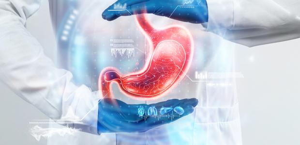
Gastroenterology is the field of medicine that specialises in the digestive tract and all its constituent organs. Gleneagles Global Health City is equipped with a distinguished team of gastroenterologists from the best medical institutions. All of our staff, from our experienced doctors and technicians to our nursing and support staff, are well-trained in their speciality and provide the highest quality patient care. We have a strong culture of community within our department, ensuring nothing is compromised when it comes to your healing.

The Institute of Advanced Gastroenterology at GGHC has the expertise to treat a gamut of gastrointestinal conditions, ranging from common disorders to rare ones. Our specialised labs and procedure rooms are equipped to perform advanced medical tests. Patients are presented with a highly competent diagnostic set-up and multiple options for treatment. They have access to world-class specialists and surgeons, caring for them round-the-clock.

Why Choose Us?

Latest Technologies

The GGHC Gastroenterology team stays updated on medical advancements and strives to provide cutting edge medical care to all our patients. We aim to usher in the future of high-quality medical care in Chennai.

best doctors india liver diseases

Patient Experience

To us, patient care is the most important criteria for a successful hospital. We focus on patient education and comfort, ensuring that they have all the information needed to make informed choices about their health. We care about how you feel from the second you walk in through our doors, until you walk out, healed.

treatment of liver diseases

Providing Quality Care

Our team believes in healing through compassion, with the strictest attention to detail. We ensure we provide all our patients with the best treatment, allowing for quick recovery. Our passion and dedication to patient care are unparalleled.

best hospital liver diseases treatment

Common Gastrointestinal Ailments

Achalasia

Achalasia Overview

Achalasia is a condition in which the muscles of the esophagus lose the ability to properly conduct food from the mouth to the stomach. Achalasia could occur when the nerves in the esophagus get damaged and the esophageal muscles may dilate or become weak. The food could get partially held back in the esophagus, and the patient may experience regurgitation.

Signs and symptoms of Achalasia

Achalasia symptoms usually start mild and worsen over time, including – inability to swallow to both solid and liquid foods, belching, regurgitation or vomiting, heartburn, intermittent chest pain, unexplained weight loss and pneumonia (from food aspiration into the lungs).

Causes and risk factors of Achalasia

The exact causes of achalasia are still not well-understood. The specific cause varies for different cases. The condition could be hereditary, due to viral infection, auto-immune response or degeneration of nerves. Esophageal cancer could cause pseudo-achalasia. Middle-aged and older patients, as well as patients with auto-immune conditions, are at a higher risk of achalasia.

Complications of Achalasia

Achalasia has no cure but the condition can be managed with medications, minimally-invasive therapies and surgical interventions if diagnosed early.

Diagnosis of Achalasia

Achalasia is usually diagnosed in one of the following ways – Esophageal manometry, X-ray or endoscopy of the esophagus, or a barium swallow test. The barium swallow test is done by asking the patient to swallow barium in a safe, liquid form and tracking its path through the esophagus using X-rays.

Treatment and Surgical Interventions of Achalasia

Achalasia in mild or early cases can be treated by a host of non-surgical options – Pneumatic dilation with a balloon, Botox or muscle relaxants. Myotomy (dividing LES) offers a high success rate and durable response to Achalasia cardia. Conventionally myotomy is performed surgically which is known as Heller’s Myotomy. A newer non-surgical, endoscopic procedure is called POEM (Per Oral Endoscopic Myotomy) is equivalent to surgical myotomy.

Prevention of Achalasia

Since the causes of achalasia are not well-known, there are no generally accepted preventive measures.

Anal Fissure

Anal fissure Overview

An anal fissure is a small tear in the thin membrane surrounding the anus known as the mucosa. An anal fissure could form while passing very hard stools. The tear could expose the anal sphincter, a ring of muscle keeping the anus closed, and cause spasms and infection. An anal fissure is considered acute if symptoms last less than 6 weeks, and could be considered chronic if symptoms last longer.

Signs and symptoms of Anal fissure

Anal fissures are characterised by extreme pain during bowel movements. The pain may last for several hours after the bowel movement too. In some cases, there is a spot of blood on the stool or the toilet paper after the bowel movement. On inspection, you may find a skin tag or a crack in the skin surrounding the anus.

Causes and risk factors of Anal fissure

Infants are quite prone to anal fissures. Anal fissures are commonly caused by constipation, very hard stools, chronic diarrhoea, childbirth or anal intercourse. The fissures could also be caused by anal cancer, HIV, Crohn’s Disease, TB or syphilis.

Complications of Anal fissure

Anal fissures sometimes extend into the anal sphincter and cause further complications while healing. If the fissure hasn’t healed properly, it will tend to get infected, cause more pain and recur frequently. An anal fissure that hasn’t healed in longer than 8 weeks is considered chronic and may require deeper interventions.

Diagnosis of Anal fissure

Often, an anal fissure can be diagnosed by a doctor with just a physical exam – A clear tear is usually visible. In case of any underlying conditions or if the doctor deems further investigation necessary, you may be prescribed a Flexible Sigmoidoscopy or a Colonoscopy.

Treatment and Surgical Interventions of Anal fissure

Anal fissures can heal on their own within a couple of weeks if efforts are taken to prevent constipation and diarrhoea. Soaking the area in warm water multiple times a day can also help relax the sphincter and promote healing. However, for persistent fissures, the doctor may recommend a course of Botox to relax the sphincter, external nitroglycerine creams, and other topical anaesthetic creams. Chronic anal fissures will require a surgical approach. The surgeon will perform a Lateral Internal Sphincterotomy (LIS), in which a small portion of the sphincter muscle is cut to reduce spasms and promote healing. Surgery is proven to be the most effective treatment for chronic anal fissures.

Prevention of Anal fissure

If you are prone to anal fissures, it is recommended to take measures to prevent constipation as well as diarrhoea. Eat high-fibre foods and stay properly hydrated. Exercise also helps, so the patient strains less during a bowel movement.

Aphthous Ulcer

Aphthous Ulcer Overview

An Aphthous Ulcer, also known as a Canker Sore, is a small round or oval sore that forms beneath the mucous membrane in the mouth or at the base of the gums. They are non-contagious but can make eating and talking quite painful. Typically, aphthous ulcers heal on their own in 7-10 days.

Signs and symptoms of Aphthous Ulcer

Most aphthous ulcers are small round or oval pustules, white or yellow in colour with a red outline. They are typically found on the insides of the cheeks, under the tongue, on the gums, or on your soft palate. They are usually extremely painful and you must seek medical help immediately if your ulcers are accompanied by a fever.

Causes and risk factors of Aphthous Ulcer

The exact cause of aphthous ulcers is uncertain, but it seems to be a combination of factors. The ulcers could be triggered by food allergies, hormonal shifts, minor mouth injury from dental work or harsh brushing, Coeliac disease, Crohn’s disease or any other auto-immune condition. Aphthous ulcers can occur in anyone but they are more common among teenagers and young adults, especially females. There also appears to be a relation to family history.

Complications of Aphthous Ulcer

Aphthous ulcers on their own are usually not serious and resolve themselves in 1-2 weeks. However, if they are acute, they could be symptoms of Crohn’s disease, Coeliac disease or an auto-immune disorder.

Diagnosis of Aphthous Ulcer

Patients affected by minor aphthous ulcers do not require tests. They can be diagnosed by a physical examination conducted by the doctor. For recurring or complex aphthous ulcers, the doctor may require a blood count, B-12 and folate studies, in addition to a Gluten Anti-body test for Coeliac disease and a Fecal Calprotectin test for Crohn’s disease.

Treatment and Surgical Interventions of Aphthous Ulcer

Minor aphthous ulcers usually don’t require any medication. With proper diet, hydration and oral hygiene, the ulcers will heal on their own in 1-2 weeks. Large or persistent ulcers will benefit from medical mouth rinses and topical products to relieve pain and speed up healing. If topical applications do not work, the doctor may prescribe oral medications to speed up healing. In extreme cases, the doctor may recommend cauterisation of the sores to reduce bleeding and reduce pain.

Prevention of Aphthous Ulcer

Aphthous Ulcers often recur so if you have a history of them, here are some precautions you can take – avoid foods you are allergic or sensitive to, drink plenty of water, follow good oral hygiene practices and practice techniques like meditation or mindfulness to reduce stress.

Autoimmune Pancreatitis

Auto-Immune Pancreatitis Overview

Auto-Immune Pancreatitis (AIP) is a chronic inflammatory condition caused by the body’s auto-immune response to the pancreas. There are currently 2 types of this condition known to medical science – Type 1 is IgG4-related pancreatitis, which affects multiple organs including the pancreas, liver, bile duct, lymph nodes, and kidneys. Type 2 is idiopathic duct-centric pancreatitis, which affects only the pancreas and is associated with inflammatory bowel disease.

Signs and symptoms of Auto-Immune Pancreatitis

AIP can often present no physical symptoms and may go undetected for years. When symptoms are present, the patient will experience nausea, weakness, loss of appetite, unexplained weight loss, jaundice, dark urine and pale stools.

Causes and risk factors of Auto-Immune Pancreatitis

As with all autoimmune conditions, it is unclear what triggers the body to attack its healthy tissue. Males over 60 are at high risk for Type 1 AIP and people above 40 with a history of Irritable Bowel Syndrome are at high risk for Type 2 AIP.

Complications of Auto-Immune Pancreatitis

AIP can cause other complications in the body such as Pancreatic stones, Diabetes and Pancreatic insufficiency.

Diagnosis of Auto-Immune Pancreatitis

To diagnose AIP, the doctor will require an IgG4 serum test, Ultrasound imaging and CAT scans of the pancreas, and in some cases a biopsy of the pancreas to rule out pancreatic cancer.

Treatment and Surgical Interventions of Auto-Immune Pancreatitis

AIP responds well to treatment with steroids. In cases with extreme jaundice, the doctor may insert a biliary stent before starting any treatment to drain the biliary duct. About 30-50% of cases will recur and require immunosuppressant treatment. The doctor will continue to monitor the liver, lymph nodes and kidneys for signs of damage as well.

Prevention of Auto-Immune Pancreatitis

The cause of AIP is unknown because of which there are no accepted preventive measures.

Clostridium Difficile Infection

Clostridium Difficile Infection Overview

Clostridium difficile is a bacteria that infects the intestine. Some patients with clostridium difficile in their system are merely carriers, never exhibiting any symptoms.

Signs and symptoms of Clostridium Difficile Infection

Mild infections usually present with symptoms like watery diarrhoea and abdominal cramping and tenderness. More severe cases may, also, experience fever, rapid heart rate, blood in the stool, increased white blood cell count and kidney failure. Extreme cases may experience intestinal inflammation, enlargement of the colon, and sepsis.

Causes and risk factors of Clostridium Difficile Infection

The infection typically affects older adults. It can also flare up due to prolonged use of antibiotics.

Complications of Clostridium Difficile Infection

Clostridium difficile is more complicated in older patients who are more prone to dehydration and kidney failure. The infection could cause toxic megacolon and bowel perforations which could turn fatal.

Diagnosis of Clostridium Difficile Infection

Clostridium difficile infection is diagnosed using a gamut of stool tests. These tests detect the presence of the Clostridium difficile toxin in the stool and hence confirm the diagnosis. The doctor may also check the status of the colon and the bowel if the infection is severe.

Treatment and Surgical Interventions of Clostridium Difficile Infection

The infection is treated with a course of antibiotics which inhibit the growth of the clostridium difficile bacteria. Other medications may also be prescribed to keep the diarrhoea, dehydration or fever under control. In critical infections, where the colon is severely damaged, surgery may be performed at the doctor’s discretion to remove the diseased portions. It is very important to finish the course of antibiotics exactly as prescribed by the doctor, as the risk of reinfection is high.

Prevention of Clostridium Difficile Infection

Clostridium difficile infections are prevented in the same way as most other bacterial infections – practice good hygiene. Wash your hands often, and especially before eating. Avoid unnecessary use of antibiotics, and follow the course exactly as prescribed by your physician.

Coeliac Disease

Coeliac Disease Overview

Coeliac disease is a serious auto-immune condition, caused by a genetic predisposition, where the patient suffers damage to the small intestine while ingesting gluten. The immune response to gluten damages the villi (finger-like projections in the small intestine that aid nutrient absorption), causing villous atrophy.

Signs and symptoms of Coeliac Disease

Coeliac disease has the following symptoms triggered by ingesting gluten – severe abdominal pain, anaemia, bloating, heartburn, constipation, diarrhoea, gas. It can also trigger a skin rash, headaches, mouth ulcers, tingling in the hands and feet and bone or joint pain.

Causes and risk factors of Coeliac Disease

The symptoms of Coeliac disease are caused when the patient ingests gluten. The cause of the condition seems to have a strong genetic link. However, there also appears to be a link between gut bacteria and infant feeding practices. People with a family history of Coeliac disease, and people with pre-existing Type-1 Diabetes, Down Syndrome, Auto-immune Thyroid disease, Lymphocytic colitis and Addison’s disease are at a higher risk of developing Coeliac disease.

Complications of Coeliac Disease

Coeliac disease can cause serious malnutrition and lactose intolerance, resulting in bone weakening, problems with the nervous system, infertility and increased risk of cancer.

Diagnosis of Coeliac Disease

Coeliac disease is diagnosed using a combination of 2 blood tests – A Serology test to check for antibodies created in response to gluten, and a DNA test to check for Genetic Predisposition to Coeliac disease. If the blood tests indicate Coeliac disease, the doctor may follow up with an endoscopy or a capsule endoscopy to review the extent of damage to the villi. In addition, the doctor may recommend a skin biopsy to check for dermatitis herpetiformis.

Treatment and Surgical Interventions of Coeliac Disease

The only way to manage Coeliac disease is a strict gluten-free diet. A licensed dietician can help create a gluten-free diet avoiding all the trace forms of gluten. Removal of gluten from the diet gradually reduce inflammation and then allow healing.

Prevention of Coeliac Disease

Coeliac disease cannot be prevented, but a flare-up can be prevented by sticking to a gluten-free diet.

Constipation

Constipation Overview

Constipation is one of the most common gastrointestinal disorders. It occurs when your bowel movements become less frequent than usual and more difficult. The stools may be very hard to pass. Constipation occurs when the colon absorbs too much water from the stools. Chronic constipation can persist for several weeks, and the patient must seek medical help immediately.

Signs and symptoms of Constipation

Constipation could make it feel like a huge strain to have a bowel movement. The stool may be very lumpy and hard and is usually very infrequent (less than 3 times a week). It could also feel as though there is a blockage in the rectum, or like you are unable to empty the rectum. You might also feel bloated, nauseous and pass a lot of gas.

Causes and risk factors of Constipation

Constipation is mostly caused by improper diet and dehydration. Stress and resisting the urge to have a bowel movement could also result in constipation. Certain medications are also known to cause constipation, and in such cases, extreme attention must be paid to following a high-fibre diet and drinking sufficient water.

Complications of Constipation

Chronic constipation can lead to haemorrhoids, anal fissures, diverticulitis or stress urinary incontinence.

Diagnosis of Constipation

The doctor will start with a medical history, a physical examination and a digital rectal examination to diagnose constipation. In addition to these, the doctor may prescribe a series of other imaging tests, blood tests or a colonoscopy to investigate the cause of constipation if needed.

Treatment and Surgical Interventions of Constipation

The first course of treatment recommended for constipation would be a series of dietary changes. A higher fibre intake, regular exercise and staying well hydrated will usually help. However, if there is no marked improvement, the doctor may prescribe laxatives or a stool softener. In extreme cases, the patient may need an enema. If the constipation is caused by a chronic obstruction in the bowel, it may be surgically corrected.

Prevention of Constipation

To prevent constipation, a high-fibre diet is recommended. Stay sufficiently hydrated and get some exercise every day.

Crohn’s Disease

Crohn’s Disease Overview

Crohn’s disease is a type of inflammatory bowel disease. It causes inflammation of various organs in the abdomen, but mainly the small intestine. Crohn’s disease can be crippling, causing abdominal pain, severe diarrhoea, weight-loss and fatigue. There is no known cure for Crohn’s disease, but modern medical therapies have been proven to greatly reduce the symptoms and even cause remission.

Signs and symptoms of Crohn’s Disease

Crohn’s disease can affect any part of the digestive tract, sometimes multiple segments. Crohn’s disease can cause abdominal pain, cramping, diarrhoea, blood in the stools, loss of appetite and unexplained weight loss, fever, fatigue and even anal fistulas. It can also cause inflammation of the skin, the eyes, or the liver and bile duct.

Causes and risk factors of Crohn’s Disease

The exact cause of Crohn’s disease is unknown. Diet and stress appear to aggravate the condition. A genetic factor is suspected to contribute to the condition but it is unsubstantiated as of now.

Complications of Crohn’s Disease

Crohn’s disease causes severe malnutrition. It can also lead to bowel obstructions, ulcers, fistulas, anal fissures, and colon cancer. Crohn’s patients also tend to have a greater risk of blood clots.

Diagnosis of Crohn’s Disease

There is no single test for Crohn’s disease. The doctor will eliminate other causes of the inflammation with a series of blood and stool tests. This may be followed by a colonoscopy, an abdominal CT, an MRI and a capsule endoscopy.

Treatment and Surgical Interventions of Crohn’s Disease

There is no known cure for Crohn’s disease and the same therapies do not work for all patients. The main goal of treatment is to reduce inflammation and limit complications. Anti-inflammatory drugs could provide symptomatic relief. Immune suppressors, biologics and antibiotics are prescribed to treat other bacteria in the gut which trigger the inflammation. The doctor may also recommend short term nutrition therapy to help to improve nutrition delivered to the body and also allow the bowel to “reset”. In very severe cases, surgery may be required to remove the damaged portions of the digestive tract.

Prevention of Crohn’s Disease

The cause of Crohn’s disease has not been identified but lifestyle and diet factors seem to affect the same. To prevent a flare-up of Crohn’s, maintain a healthy diet, practice stress-relieving activities and avoid smoking.

Diverticulitis

Diverticulitis Overview

Diverticula are small bulging pouches that sometimes form in the intestinal lining. When those pouches get infected, the resulting condition is Diverticulitis. Diverticula are common in people above 40, and usually don’t cause major problems. However, when they get infected, they cause abdominal pain, swelling, and fever.

Signs and symptoms of Diverticulitis

Diverticulitis can cause abdominal pain, blood in the stools, bloating, nausea, and constipation.

Causes and risk factors of Diverticulitis

Diverticulitis usually occurs when the diverticula tear and get infected, or get blocked with stool. This is most common in people above the age of 40. People who are overweight, smoke cigarettes, lead sedentary lives and have insufficient fibre in their diet are also at an increased risk of diverticulitis.

Complications of Diverticulitis

If left untreated, diverticulitis can cause further complications such as abscesses around the diverticula, intestinal perforations, scarring and fistulas.

Diagnosis of Diverticulitis

The doctor will begin with a physical exam, and then a series of blood tests, urine tests and stool tests, to find the infection. The doctor will then perform a CT scan to look for infected and inflamed diverticula. A liver enzyme test may also be needed.

Treatment and Surgical Interventions of Diverticulitis

If the diverticulitis is mild, the doctor may recommend antibiotics along with rest and plenty of fluids to allow the walls of the intestine to heal. For more serious cases, intravenous antibiotics and fluids may be required, necessitating a hospital stay. If there is an abscess, the doctor will drain it. In extreme cases, where the intestine has ruptured, the doctor may opt to go for a surgical repair. Once healed, the doctor may perform a colonoscopy to rule out colon cancer.

Prevention of Diverticulitis

To prevent diverticulitis, optimise your fibre-intake, exercise regularly and hydrate. If you are a smoker, stop.

Dysphagia

Dysphagia Overview

Dysphagia is a condition where the patient has difficulty to swallow either water, food or both. Swallowing is a complex activity involving the coordinated functioning of multiple involuntary muscles. This coordinated action is called peristalsis. When the co-ordination gets thrown off, it causes dysphagia.

Signs and symptoms of Dysphagia

Dysphagia may present as difficulty swallowing, frequent choking on food or water, pain while swallowing and frequent pneumonia due to food entering the wind-pipe. If left unnoticed, the problem could advance and cause the patient to choke on their food.

Causes and risk factors of Dysphagia

Most common causes are cricopharyngeal web, corrosive or a peptic stricture and malignancy Dysphagia may also due to a neurological cause. It is typically present in geriatric adults. Patients with a history of other neurological disorders like Parkinson’s disease are at a higher risk for dysphagia.

Complications of Dysphagia

Dysphagia, if left untreated, could result in serious malnutrition and dehydration.

Diagnosis of Dysphagia

The diagnosis will begin with patient history and physical exam. A speech-language pathologist could be consulted to identify which muscle group is failing. A swallow study, a barium swallow test, an endoscopy and manometry test may be required to identify the extent and cause of the dysphagia.

Treatment and Surgical Interventions of Dysphagia

Because of its neurological cause, treatment of dysphagia can be tricky. For low dysphagia, the esophagus may be widened using a balloon pump or a Botox injection to relax the muscles. For high dysphagia, the patient may have to be put on a temporary feeding tube, while also undergoing speech therapy to help them learn new ways to swallow correctly. If the dysphagia is caused by cancer, the patient will be referred to an oncologist for further treatment of the cancer.

Prevention of Dysphagia

Although dysphagia can’t be prevented, the risk can be reduced by eating slowly and chewing your food well. Early detection and effective treatment of GastrEsophageal Reflux (GERD) can reduce the risk of dysphagia.

Esophageal Cancer

Esophageal cancer Overview

Esophageal cancer affects the “food pipe” which connects the mouth and the stomach. It typically starts in the lining of the esophagus and then penetrates the walls. The cancer grows and creates an obstruction in the esophagus resulting in complications, practical difficulty with swallowing etc. The most common types of Esophageal cancer are adenocarcinoma and squamous cell carcinoma.

Signs and symptoms of Esophageal cancer

Early on, there may be no clear symptoms. As the cancer advances, it may cause unexplained weight loss, fatigue, pain while swallowing, chest pain, coughing, indigestion and heartburn.

Causes and risk factors of Esophageal cancer

The cause of Esophageal cancer is not clear. However, smoking, heavy alcohol abuse and a history of GERD are linked to a higher risk of Esophageal cancer. Elderly, obese men are a particularly high-risk demographic.

Complications of Esophageal cancer

Esophageal cancer could grow and cause an obstruction. It could also cause internal bleeding and severe pain.

Diagnosis of Esophageal cancer

Esophageal cancer is confirmed with a series of imaging and diagnostic tests, including endoscopy and biopsy.

Treatment and Surgical Interventions of Esophageal cancer

The treatment plan will be formulated for each patient depending on the extent of the cancer, the type of cell that has caused the cancer, and the baseline health of the patient. Chemotherapy and radiation therapy could benefit the patient. Additional surgical intervention may or may not be required. Targeted-drug therapy and immunotherapy have also shown results in the treatment of Esophageal cancer. Endoscopy guided Esophageal stent placement (SEMS) helps to relieve difficulty in swallowing.

Prevention of Esophageal cancer

To reduce your risk of Esophageal cancer, quit drinking and smoking, eat healthier and cleaner and maintain a good Body Mass Index (BMI).

Esophageal Spasms

Esophageal Spasms Overview

Esophageal spasms are painful contractions of the esophagus. It can feel like spontaneous chest pains that last anywhere from 5 mins to a couple of hours. They occur rarely and usually don’t require any treatment. But if the spasms occur often, they may require medical attention.

Signs and symptoms of Esophageal Spasms

Esophageal spasms can be mistaken for heart pain. It usually presents as difficulty swallowing, regurgitation or the feeling of an obstruction in the esophagus. The spasms may be triggered by hot or cold foods and liquids.

Causes and risk factors of Esophageal Spasms

Medical science is unsure what causes these spasms but they appear to be neurological. There are 2 types of esophageal spasms based on the muscle movement involved – Distal Esophageal spasms (DES) involving uncoordinated “up-and-down” spasms, or Jack-hammer esophageal spasm which involves a corkscrew movement resulting in more intense spasms. Esophageal spasms are more likely in geriatric patients with GERD, high blood pressure and anxiety or depression.

Complications of Esophageal Spasms

Esophageal spasms could cause chest pains and reduce the quality of life. In the case of pre-existing injury or perforation in the esophagus, the spasms could cause further tearing of the tissue.

Diagnosis of Esophageal Spasms

The doctor will first rule out a cardiac condition by performing an EKG. Then, an esophageal manometry test and barium swallow test are needed to check the function of the esophageal muscles. An endoscopy may be required. The doctor may also see the need to monitor the pH of the esophagus to check for acid reflux.

Treatment and Surgical Interventions of Esophageal Spasms

To treat the esophageal spasm, it is essential to identify the cause. If certain foods or certain temperatures are triggers, these must be eliminated from the diet. Quitting smoking, maintaining a healthy weight and eating smaller, more frequent meals could help. Botox injections could help patients by relaxing theesophageal muscles. Surgery is recommended very rarely. A Peroral Endoscopic Myotomy (POEM) is performed to surgically remove a portion of the esophagus to reduce contraction.

Prevention of Esophageal Spasms

Identifying and eliminating the triggers can help prevent recurrent bursts of esophageal spasms. Controlling underlying conditions that cause the spasms, such as depression or GERD, will also help.

Esophageal Ulcer

Esophageal Ulcer Overview

Esophageal ulcers are peptic ulcers in the membrane of the lower portion of the esophagus. They occur when the mucous membrane of the esophagus thins down, making the lining vulnerable to infection or damage. These ulcers can be caused by acid reflux from the stomach. They are also caused by infections, from a range of bacteria, yeasts and viruses.

Signs and symptoms of Esophageal Ulcer

Esophageal ulcers usually feel like a burning pain below the sternum, in the centre of the chest. The patient may also experience heartburn, nausea, shortness of breath, difficulty swallowing, acute abdominal pain, vomiting and unexplained weight-loss.

Causes and risk factors of Esophageal Ulcer

Esophageal ulcers are usually caused due to damage from stomach acids. They dissolve the mucous membrane and attack the esophageal lining. This becomes a point of potential infection. Certain medications can also have this effect. Alcohol abuse can also alter stomach chemistry and cause Esophageal ulcers.

Complications of Esophageal Ulcer

The main complication associated with ulcers is recurrence. It could also cause gastrointestinal bleeding and perforation.

Diagnosis of Esophageal Ulcer

The diagnosis for an esophageal ulcer is based on the medical history, physical exam and an endoscopy to look inside the esophagus. A barium X-ray may also be required. After the ulcer is diagnosed, the doctor may take a sample of the ulcer tissue for further analysis.

Treatment and Surgical Interventions of Esophageal Ulcer

The treatment of the ulcer depends on the identified cause. If the ulcer is bacterial, the doctor may prescribe antibiotics. If the cause is another medication for your pre-existing conditions, the doctor may change that course of treatment. You may also be prescribed over-the-counter H2 receptor blockers to reduce stomach acid production. The doctor may also prescribe a proton pump inhibitor to allow the esophageal wall to heal.

Prevention of Esophageal Ulcer

Esophageal ulcers can be prevented by drinking adequate water, eating small, frequent meals, taking precautions to prevent acid reflux and bacterial gut infections.

Esophageal Varices

Esophageal Varices Overview

Esophageal varices are enlarged veins in the esophagus, commonly occurring in patients with liver disease. The varices can be asymptomatic if mild but may turn dangerous if they rupture and bleed.

Signs and symptoms of Esophageal Varices

Most patients do not realise they have varices until they rupture, as they are largely asymptomatic. When they rupture, the patient may vomit blood, have bloody stools, experience symptoms of shock and faint.

Causes and risk factors of Esophageal Varices

Esophageal varices usually occur in patients with liver disease. Due to disease, the blood pressure in the portal vein increases and pushes blood into the surrounding organs, like the esophagus. This additional pressure creates swelling in the veins and then forms varices. If the blood pressure exceeds the threshold amount, the patient experiences rupture. Liver cirrhosis, fatty liver disease or viral hepatitis could cause this condition. Patients with high blood pressure, alcoholics and patients in liver failure are at a higher risk of esophageal varices rupturing.

Complications of Esophageal Varices

The most serious complication from esophageal varices is bleeding. If the body loses too much blood, it can go into shock and start shutting down.

Diagnosis of Esophageal Varices

Patients with liver disease should be screened for esophageal varices regularly. This is done by performing an endoscopy. An additional CT scan or MRI may also be required.

Treatment and Surgical Interventions of Esophageal Varices

Treatment for Esophageal varices targets 3 things – prevention of further liver damage, prevention of varices rupturing and control of bleeding in case of rupture. Further liver damage is prevented by avoiding the consumption of food or beverages that build up toxins in the body. The aim is to have a diet that gives the liver a break. The doctor will also prescribe medication to keep the blood pressure down. This will help prevent the varices from rupturing. In case of rupture, the doctor will control and prevent bleeding through the application of pressure and various pressure-reducing medications. The liver condition will have to be treated aggressively and in extreme cases, a liver transplant may be required.

Prevention of Esophageal Varices

Keep your liver healthy. Reduce intake of alcohol and fatty foods. Keeping your blood pressure in the recommended range will also help.

Esophagitis

Esophagitis Overview

Esophagitis is an inflammation in the lining of the esophagus. If untreated, this can cause discomfort to the patient and lead to alterations in the structure of the esophagus.

Signs and symptoms of Esophagitis

Esophagitis is most commonly associated with abdominal pain, nausea, heartburn, mouth sores, lack of appetite and unexplained weight-loss.

Causes and risk factors of Esophagitis

Esophagitis can be caused by a variety of factors – GERD, Allergies, infections, ingestion of chemical or certain medication. Smoking, pregnancy and a weak immune system increase the risk of esophagitis.

Complications of Esophagitis

Left untreated, esophagitis could cause scarring and narrowing of the esophagus, tearing of the esophagus and increase your risk of esophageal cancer.

Diagnosis of Esophagitis

In addition to the patient medical history and a physical exam, the doctor may call for a Barium X-ray, an endoscopy and an allergy panel. A small tissue sample may also be taken to test for pre-cancer cells.

Treatment and Surgical Interventions of Esophagitis

Treatment of esophagitis is intended to manage inflammation, treat complications and address the underlying cause of the condition. The doctor may prescribe antacids, H2 receptor blockers, proton pump inhibitors, steroidal medication or any other drug relevant to the cause of the inflammation.

Prevention of Esophagitis

Preventing acid reflux, keeping your allergies in check and avoiding certain medications can help reduce the risk of esophagitis.

Fecal Incontinence

Fecal Incontinence Overview

Fecal incontinence occurs when the patient is unable to hold or control their bowel movements. Common causes of fecal incontinence include diarrhoea, constipation, and muscle or nerve damage. This may be due to ageing or giving birth. Although it may be embarrassing to talk about, please seek medical attention to improve your quality of life. We assure you that our doctors are sensitive to your emotions and will do their best to help you get better.

Signs and symptoms of Fecal Incontinence

Fecal incontinence may occur temporarily during a severe round of diarrhoea and subside once the infection has passed. However, it can also turn into a chronic issue. It is usually accompanied by gas, bloating and diarrhoea.

Causes and risk factors of Fecal Incontinence

There can be various reasons for fecal incontinence to occur – infection and diarrhoea, damage to the anal sphincter, nerve damage, constipation (which leads to nerve damage), rectal prolapse or haemorrhoids. Fecal incontinence is more common in geriatric patients, with pre-existing conditions like dementia or Parkinson’s disease. Patients who have diabetes, multiple-sclerosis or back trauma are also at a higher risk for fecal incontinence.

Complications of Fecal Incontinence

Fecal incontinence can cause serious skin rashes around the anus and risk of infection, not to mention serious emotional distress to the patient.

Diagnosis of Fecal Incontinence

Fecal incontinence is usually diagnosed with patient history and a physical exam. The doctor may also want a Digital Rectal Exam, Anal Manometry, a Colonoscopy and an MRI.

Treatment and Surgical Interventions of Fecal Incontinence

Depending on the cause, the doctor may prescribe anti-diarrhoea drugs or laxatives. The doctor may also recommend practising Kegels, Bowel Training exercises, Sacral Nerve Stimulation (SNS) Therapy, and Radiofrequency Therapy. If there is sphincter damage caused due to rectal prolapse or childbirth, the doctor may opt for a surgical fix like a Sphincteroplasty. As a last resort, the doctor may opt for the insertion of a Colostomy bag.

Prevention of Fecal Incontinence

Depending on the cause, reducing constipation as well as diarrhoea, having a healthy amount of fibre in the diet and staying hydrated will help prevent fecal incontinence.

Gastritis

Gastritis Overview

Gastritis is a condition in which the lining of the stomach gets inflamed. The inflammation is usually caused by a bacterial infection and may lead to stomach ulcers too. Acute gastritis occurs suddenly and with intense symptoms. Chronic gastritis occurs gradually, the intensity of the symptoms worsening over weeks.

Signs and symptoms of Gastritis

Gastritis presents as a burning or gnawing pain, like indigestion, nausea, vomiting and bloating in the upper abdomen.

Causes and risk factors of Gastritis

Gastritis occurs when the mucous lining of the stomach is weakened due to caustic stomach acids. Several pre-existing digestive disorders like Crohn’s disease can accelerate gastritis. Bile reflux and bacterial infections can also cause gastritis. Older adults, alcoholics, very stressed people and patients with auto-immune conditions have a greater risk of gastritis.

Complications of Gastritis

Gastritis, if left untreated, can cause bleeding and stomach ulcers. In rare cases, chronic gastritis increases the chances of stomach cancer.

Diagnosis of Gastritis

Gastritis is diagnosed based on the patient’s medical history, physical exam, an endoscopy to inspect the upper digestive tract and a Barium X-ray. The doctor may also test for bacteria H.Pylori.

Treatment and Surgical Interventions of Gastritis

Gastritis is treated with antibiotics, antacids, proton pump inhibitors or Histamine (H2) blockers or a combination of them.

Prevention of Gastritis

Gastritis can be prevented by avoiding spicy, fatty or fried foods, alcohol and smoking. Decreasing stress and avoiding unnecessary medications can also be helpful.

Gastroenteritis

Gastroenteritis Overview

Gastroenteritis is stomach flu characterised by watery diarrhoea and intense abdominal pain. It is usually caused by a bacterial or viral infection transmitted via food or water. Gastroenteritis is common in both children and adults.

Signs and symptoms of Gastroenteritis

Gastroenteritis presents as watery diarrhoea, abdominal pain and cramps, nausea, vomiting, bloating and sometimes fever.

Causes and risk factors of Gastroenteritis

Gastroenteritis is usually caused by a bacteria or virus contaminating the food or water consumed by the patient. It could also be a result of sub-par hand-washing practices. Infants and geriatric patients are especially vulnerable to Gastroenteritis. Patients with weakened immune systems are also at risk.

Complications of Gastroenteritis

The risk of dehydration is the main complication to watch out for while suffering from gastroenteritis. Be sure to replace fluids and salts lost in vomiting and diarrhoea. In extreme cases, intravenous hydration may be necessary.

Diagnosis of Gastroenteritis

Gastroenteritis is diagnosed based on symptoms, patient medical history and a physical exam. If it is necessary to identify the cause of infection, the doctor may prescribe a stool sample test.

Treatment and Surgical Interventions of Gastroenteritis

There is no treatment for viral gastroenteritis, so rest and fluids are recommended. For bacterial gastroenteritis, the doctor may prescribe antibiotics and also some general probiotics and vitamins.

Prevention of Gastroenteritis

The best way to prevent the contraction of gastroenteritis is to vaccinate and practice good hygiene. This is more critical in public or shared spaces and while travelling.

Gastroesophageal Reflux (GERD)

Gastroesophageal Reflux Overview

GERD occurs when stomach acid refluxes into the esophagus, past the Lower Esophageal Sphincter (LES) and irritates the mucous membrane and inner lining. If you suffer from GERD, you might experience it as heartburn or acid indigestion. Acid reflux is common, but when it turns chronic (at least twice a week, of moderate-intensity), it could be GERD and the patient must seek help.

Signs and symptoms of Gastroesophageal Reflux

Common symptoms of GERD are – heartburn, chest pain, difficulty swallowing, vomiting, laryngitis and disturbed sleep.

Causes and risk factors of Gastroesophageal Reflux

GERD is a chronic acid reflux condition caused by the inefficiency of the LES. The sphincter doesn’t close completely, allowing acid backflow the consequent symptoms. Patients with Hiatal hernias, weight issues and connective tissue disorders like scleronoma are particularly at risk for GERD. Smoking, eating large meals at odd times, coffee, alcohol and certain medications like aspirin could increase your risk of acid reflux and hence GERD.

Complications of Gastroesophageal Reflux

Chronic GERD can cause ulcers in the esophagus, obstructions or narrowing of the esophagus, and Barrett’s esophagus (pre-cancerous changes).

Diagnosis of Gastroesophageal Reflux

GERD is diagnosed based on the patient’s medical history, a physical exam, an upper digestive tract endoscopy, a pH test of the lower esophagus, an X-ray and an esophageal manometry test to measure muscular contraction.

Treatment and Surgical Interventions of Gastroesophageal Reflux

If mild, doctors may first recommend that you try lifestyle changes and prescribe over-the-counter antacids, H2 receptor blockers or proton pump inhibitors. For chronic cases that do not experience relief in a few weeks, stronger versions of the over-the-counter medication may be prescribed. However, if the medication does not make GERD manageable, the patient will require surgery to correct the LES. Depending on the case, the doctor may recommend a Fundoplication, a LINX device, or a Transoral Incision less Fundoplication.

Prevention of Gastroesophageal Reflux

GERD can be prevented by maintaining a healthy weight, avoiding foods that trigger reflux, quitting smoking and sleeping with the head elevated (for night-time reflux).

Gastrointestinal Bleeding

Gastrointestinal (GI) bleeding Overview

Gastrointestinal (GI) bleeding is a serious symptom that something is wrong in the digestive tract. It could be anywhere from the inside of the mouth to the anus. The amount of bleeding can also vary from minor to fatal. You can usually tell you have experienced gastrointestinal bleeding by the presence of blood in your vomit or stools.

Signs and symptoms of Gastrointestinal (GI) bleeding

Gastrointestinal bleeding usually presents as blood in the stool or vomit, but sometimes, the blood could also be a black tarry substance. If it is serious, it is usually accompanied by weakness, dizziness, clammy and pale skin and shortness of breath.

Causes and risk factors of Gastrointestinal (GI) bleeding

Gastrointestinal bleeding can occur in the upper or lower digestive tract and due to a large variety of causes. It could occur due to peptic ulcers, varices, esophagitis, diverticulitis or haemorrhoids.

Complications of Gastrointestinal (GI) bleeding

Severe gastrointestinal bleeding could cause shock, anaemia or even death.

Diagnosis of Gastrointestinal (GI) bleeding

The diagnosis for gastrointestinal bleeding is done based on a series of blood tests, stool tests, an endoscopy, an upper gastrointestinal lavage, computed tomography abdominal scan and possibly a flexible sigmoidoscopy.

Treatment and Surgical Interventions of Gastrointestinal (GI) bleeding

Often GI bleeding stops on its own. However, if it doesn’t, the cause of the bleeding must be identified and then treated accordingly. Usually, a proton pump inhibitor is prescribed to suppress stomach acid production. If the bleeding is very severe, the patient may also require a blood transfusion and Non-steroidal anti-inflammatory drugs (NSAIDs) or Aspirin.

Prevention of Gastrointestinal (GI) bleeding

To help prevent GI bleeding, limit the use of cigarettes, alcohol and NSAIDs. If you have pre-existing GERD, make sure to keep that under control.

Haemorrhoids

Haemorrhoids Overview

Haemorrhoids or piles are swollen veins in the rectum, similar to varicose veins. Haemorrhoids can also develop in the skin around the anus. They are common, and nearly 3 out of 4 adults will develop haemorrhoids in their lifetime. Haemorrhoids are the most common cause of rectal bleeding.

Signs and symptoms of Haemorrhoids

Haemorrhoids are of 2 types – internal and external. Internal haemorrhoids are usually not visible and usually hurt less. You would notice blood in the stools though, and in case of prolapse, the prolapsed tissue could project out and cause major pain during bowel movements. External haemorrhoids occur under the skin around your anus and cause pain, itching, bleeding and swelling.

Causes and risk factors of Haemorrhoids

Haemorrhoids can be caused due to straining during bowel movements, chronic diarrhoea or constipation, anal intercourse, a low fibre diet, and regular heavy lifting. Old age and pregnancy can also increase your risk of haemorrhoids.

Complications of Haemorrhoids

Haemorrhoids could lead to anaemia or blood clots. In rare cases, the haemorrhoid could become strangulated, i.e. have its blood supply cut off, leading to severe pain.

Diagnosis of Haemorrhoids

External haemorrhoids can be diagnosed with a physical exam and a patient medical history and maybe a digital rectal exam. However, to diagnose internal haemorrhoids, the doctor may need additional tests such as an endoscopy, a colonoscopy and a sigmoidoscopy.

Treatment and Surgical Interventions of Haemorrhoids

Basic lifestyle changes can help alleviate the symptoms of mild haemorrhoids. A fibre-full diet, adequate water and ice packs for the pain will help. The doctor will prescribe topical creams and ointments or oral painkillers for relief from itching, pain and swelling. However, for serious, chronic haemorrhoids, the doctor may choose to perform a non-invasive or surgical procedure for removal. Non-invasive procedure for removal, like a rubber-band ligation, sclerotherapy or coagulation with a laser or infrared radiation. Surgical procedures to treat haemorrhoids include haemorrhoidectomy and haemorrhoid stapling.

Prevention of Haemorrhoids

To prevent haemorrhoids, the stools need to be kept soft. Eating high fibre foods and drinking adequate water will help with this. It is also recommended to exercise and avoid long periods of sitting.

Indigestion, Non-ulcer Dyspepsia

Indigestion, Non-ulcer Dyspepsia Overview

Non-ulcer dyspepsia is a general term for recurring symptoms of indigestion, with no clear cause. Indigestion is very common in people of all ages. Typically, patients complain of pain and bloating in the upper abdomen.

Signs and symptoms of Indigestion, Non-ulcer Dyspepsia

Indigestion usually feels like a burning pain in the upper abdomen, mostly accompanied by bloating, belching and nausea. If you experience any shortness of breath, bloody vomit or tarry stools, please consult a doctor immediately.

Causes and risk factors of Indigestion, Non-ulcer Dyspepsia

The cause of non-ulcer dyspepsia is not clear. The risk of non-ulcer dyspepsia is higher in geriatric patients, women in general, patients with a history of anxiety or depression and smokers.

Complications of Indigestion, Non-ulcer Dyspepsia

The condition can cause further deterioration of the patient’s mental health.

Diagnosis of Indigestion, Non-ulcer Dyspepsia

The doctor may take a series of blood and stool tests to rule out other gastrointestinal conditions. An upper digestive tract endoscopy may also be required.

Treatment and Surgical Interventions of Indigestion, Non-ulcer Dyspepsia

Usually, a simple diet and lifestyle modifications will help ease the symptoms and the frequency of non-ulcer dyspepsia. Over-the-counter Antacids can be effective in treating indigestion. However, if chronic, the doctor may test and treat for H. pylori infections in the stomach. Proton pump inhibitors (acid-suppressing medication) may also be prescribed.

Prevention of Indigestion, Non-ulcer Dyspepsia

Indigestion can be prevented by controlling your diet – avoid excessively fatty or fried foods, chew meals well and eat 6 small meals a day. Taking up meditation or yoga to reduce day-to-day stress is also recommended.

Inflammatory Bowel Disease

Inflammatory Bowel Disease Overview

Inflammatory Bowel Disease (IBD) is an umbrella term to define conditions associated with inflammation of the intestines. While it is often assumed to be an auto-immune condition, it could also be caused due to the immune system attacking pathogens present in the intestines, thereby causing inflammation. Ulcerative Colitis and Crohn’s disease are the two major types of IBD. IBD can be debilitating and requires serious medical attention.

Signs and symptoms of Inflammatory Bowel Disease

Patients with IBD usually experience diarrhoea, blood in the stools, abdominal pain and cramping, fatigue, reduced appetite and unexplained weight-loss. The intensity of these symptoms can vary.

Causes and risk factors of Inflammatory Bowel Disease

The exact cause of IBD is not clear. It may be caused due to an auto-immune response or due to infection. Smoking, a family history of IBD and consuming NSAIDs increases your risk of developing the condition.

Complications of Inflammatory Bowel Disease

IBD increases your risk of colon cancer. It can also increase your risk of other inflammatory conditions in the body, like arthritis.

Diagnosis of Inflammatory Bowel Disease

The doctor will probably need to rule out other conditions that cause the same symptoms to diagnose IBD. This would require a panel of blood and stool tests, CT scans, X-ray scans, an endoscopy and a sigmoidoscopy. A balloon-assisted enteroscopy may also be required to confirm the diagnosis.

Treatment and Surgical Interventions of Inflammatory Bowel Disease

Treatment aims to reduce inflammation, tackle possible complications and hopefully push the condition into long-term remission. The doctor will treat the condition with a combination of anti-inflammatory drugs, immunosuppressors, biologics, antibiotics and nutritional supplements. If diet, lifestyle changes and drug therapies do not work, the doctor may choose to take a surgical approach, which varies depending on the type of IBD.

Prevention of Inflammatory Bowel Disease

IBD cannot be prevented but the risk can be significantly reduced by maintaining a healthy diet and level of hydration and quitting smoking.

Intestinal Ischemia

Intestinal Ischemia Overview

Intestinal ischemia describes a series of conditions caused due to inadequate blood supply to the intestines. Usually, this is caused by a blockage in the artery. It may affect your small intestine, large intestine or both.

Signs and symptoms of Intestinal Ischemia

Intestinal ischemia presents as abdominal cramps, abdominal pain that gets progressively worse over weeks, diarrhoea, bloating, nausea and unexplained weight-loss.

Causes and risk factors of Intestinal Ischemia

Intestinal ischemia is caused when a major artery supplying blood to the intestines develops an obstruction. Over time this obstruction could develop into a blockage. This obstruction could be due to a blood clot, cholesterol deposits etc. Pre-existing conditions like atherosclerosis, high blood pressure and congestive heart failure could increase your risk of intestinal ischemia. Consumption of cocaine and methamphetamines are also proven to increase your risk of ischemia.

Complications of Intestinal Ischemia

Complications of intestinal ischemia include gangrene, perforation of the intestinal walls, scar formation and in very extreme cases, death.

Diagnosis of Intestinal Ischemia

Intestinal ischemia is diagnosed with the aid of patient medical history, a physical exam, blood tests, X-rays, CT scans, an endoscopy and possibly an angiograph to test for arterial blockages. In some cases, exploratory surgery may also be needed to find and collect damaged tissue for further pathological studies.

Treatment and Surgical Interventions of Intestinal Ischemia

Depending on where the ischemia has occurred in the intestines, different treatment approaches are adopted. Colon ischemia usually heals on its own, but in severe cases, the damaged tissue is surgically removed. Acute mesenteric artery ischemia usually requires by-pass surgery or an angioplasty to restore blood flow to the affected areas.

Prevention of Intestinal Ischemia

The risk of intestinal ischemia can be reduced by incorporating some lifestyle changes including – quitting smoking, exercising regularly, following a healthy diet, reducing stress and controlling diabetes (if applicable).

Irritable Bowel Syndrome

Irritable Bowel Syndrome Overview

Irritable Bowel Syndrome (IBS) is a common disorder that affects the large intestine. It is unrelated to IBD and is characterised by serious tummy discomfort or pain and difficult bowel habits. There are 4 types of IBS – IBS with constipation (IBS – C). IBD with Diarrhoea (IBS – D), IBS with a combination of constipation and diarrhoea (IBS – M), and IBS – U for patients who don’t fall into the first 3 categories. IBS doesn’t increase your likelihood of colon cancer or other colon conditions, but it can seriously affect your quality of life.

Signs and symptoms of Irritable Bowel Syndrome

IBS symptoms usually include diarrhoea, constipation, abdominal cramps, gas and bloating, mucous in your stools, heartburn and a feeling of incontinence.

Causes and risk factors of Irritable Bowel Syndrome

The cause of IBS isn’t clear, but medical science has identified the triggers – stress, smoking, foods containing lactose, gluten, caffeine or carbonation. IBS occurs more commonly in people under the age of 50, especially women.

Complications of Irritable Bowel Syndrome

Chronic IBS can lead to haemorrhoids. It has also been shown to adversely affect the patient’s quality of life and performance, in addition to their mental health.

Diagnosis of Irritable Bowel Syndrome

There is no definitive test to diagnose IBS. The doctor will require a complete medical history, a physical exam and tests to rule out other conditions like Coeliac disease, lactose intolerance, or infection. The patient usually needs to have experienced abdominal pain once a week for the last 3 months, experience pain during bowel movements and have altered consistency of stools.

Treatment and Surgical Interventions of Irritable Bowel Syndrome

IBS can often be controlled by managing stress and diet factors. The doctor may also suggest laxatives, anti-diarrhoea drugs, or antacids to manage immediate symptoms. Fecal Microbiota Transplantation (FMT) is an upcoming treatment plan for IBS, currently in clinical trials.

Prevention of Irritable Bowel Syndrome

IBS is linked to stress and diet, so keeping these factors under control will reduce your risk. Practice meditation or mindfulness to relieve anxiety and stress, and be sure to take your anti-anxiety medication or anti-depressants if applicable. Following a clean and healthy diet with a good amount of fibre will also help. Reduce intake of alcoholic and carbonated beverages.

Lactose Intolerance

Lactose Intolerance Overview

Lactose intolerance is a genetic predisposition, wherein the patient is unable to digest lactose, the main carbohydrate found in dairy products. The condition is not harmful but may be uncomfortable and embarrassing. There is no cure for this condition, but it can be managed by limiting or eliminating dairy products from the patient’s diet.

Signs and symptoms of Lactose Intolerance

The symptoms of lactose intolerance usually begin about 30-60 mins after ingesting dairy products. They include bloating, diarrhoea, stomach cramps, vomiting and gas.

Causes and risk factors of Lactose Intolerance

Lactose intolerance occurs when the small intestine doesn’t produce enough lactase to digest lactose in dairy. This undigested lactose moves to the colon and interacts with the bacteria causing uncomfortable symptoms. Lactose intolerance can be present in infancy, or it may be developed in adulthood. Premature babies and patients with Crohn’s or Coeliac disease are at a higher risk of lactose intolerance.

Complications of Lactose Intolerance

Lactose intolerance in infants, if left unaddressed, can lead to malnutrition.

Diagnosis of Lactose Intolerance

The doctor could diagnose lactose intolerance based on the patient medical history, symptoms and the patient’s response to eliminating dairy from their diet. The doctor may choose to verify the diagnosis with a Hydrogen Breath test or a Lactose tolerance test.

Treatment and Surgical Interventions of Lactose Intolerance

There is no cure for lactose intolerance. If it is caused by another underlying condition, treatment of that condition could help. Otherwise, the patient is advised to avoid lactose in their diet.

Prevention of Lactose Intolerance

Lactose intolerance cannot be prevented.

Traveller’s Diarrhoea

Traveller’s Diarrhoea Overview

Traveller’s diarrhoea is an infection contracted as a result of consuming contaminated food or water. The patient usually experiences nausea, diarrhoea and abdominal cramps. Thankfully, it is not a serious condition. If you are travelling to high-risk areas, it is good to consult a doctor before-hand and take appropriate precautions.

Signs and symptoms of Traveller’s Diarrhoea

Travellers diarrhoea usually begins on a trip or shortly after. Most cases experience abrupt diarrhoea, abdominal cramps, vomiting and in some cases fever.

Causes and risk factors of Traveller’s Diarrhoea

Traveller’s diarrhoea is caused by eating or drinking something contaminated.

Complications of Traveller’s Diarrhoea

Traveller’s diarrhoea can cause dehydration. This is dangerous in children and older adults.

Diagnosis of Traveller’s Diarrhoea

Typically traveller’s diarrhoea resolves on its own within 3-4 days, but if it does not, a visit to the doctor may be required. The doctor will need a detailed patient history (be sure to mention your recent travels), a physical exam and a panel of blood and stool tests to check for infections and dehydration.

Treatment and Surgical Interventions of Traveller’s Diarrhoea

Travellers diarrhoea usually resolves itself. The diarrhoea is usually not stopped medically as its purpose is to remove the infection from the body. In case of extreme dehydration though, the doctor may prescribe antimotility agents and an intravenous IV. If you have a bacterial or parasitic infection, the doctor will prescribe antibiotics or anti-parasitic drugs accordingly.

Prevention of Traveller’s Diarrhoea

Traveller’s diarrhoea is an infection and hence can be prevented by taking appropriate measures to maintain hygiene. Avoid eating from unhygienic restaurants and carry clean water with you. Remember to wash your hands often, especially before a meal.

Ulcerative Colitis

Ulcerative Colitis Overview

Ulcerative colitis is an inflammatory bowel disease causing irritation and ulcers in the lining of the colon. It is an auto-immune disease. The condition has no cure, and patients usually have symptoms from time to time. However, with the right medical guidance, the patient can have a good quality of life.

Signs and symptoms of Ulcerative Colitis

The main symptom of ulcerative colitis is bloody stools and some occasional pus. The patient may experience sudden urges to defecate, but then not be able to do so despite the urge. Other symptoms include abdominal cramps, loss of appetite, anaemia, pain associated with bowel movements, fatigue, fever and dehydration.

Causes and risk factors of Ulcerative Colitis

Ulcerative colitis is an auto-immune disease. Its exact cause is unknown, but it occurs when the body’s immune system attacks the colon. Hereditary factors appear to have a role to play.

Complications of Ulcerative Colitis

Ulcerative colitis could cause severe internal bleeding, perforations in the colon, toxic megacolon and an increased risk of colon cancer.

Diagnosis of Ulcerative Colitis

Ulcerative colitis looks similar to other conditions like Crohn’s disease so the doctor will perform tests to rule those out. A panel of blood and stool tests, along with an endoscopy, an abdominal CT and a colonoscopy would be required to diagnose Ulcerative colitis. The doctor may also need a colon biopsy.

Treatment and Surgical Interventions of Ulcerative Colitis

Mild to moderate patients will require only drug therapy to treat their condition. This usually involves a combination of anti-inflammatory drugs, immunosuppressants, biologics and pain medication. Serious cases will require surgical removal of the colon and the rectum, known as a proctocolectomy.

Prevention of Ulcerative Colitis

Ulcerative colitis cannot be prevented.

Procedures

Endoscopic Procedures

The Department of Gastroenterology at GGHC provides specialised diagnostic and treatment options for a wide variety of gastroenterological conditions. These procedures could be diagnostic or therapeutic, depending on the disease. Our endoscopy suites are equipped with state-of-the-art equipment for fast and accurate diagnosis, clinical research and good patient care.

Diagnostic

Gastroscopy

Gastroscopy is a procedure performed using an endoscope to examine the upper GI tract (the esophagus, stomach and duodenum), especially its inner lining.

Colonoscopy

Colonoscopy is a procedure performed to detect changes or abnormalities in the rectum, colon and large intestine using an endoscope.

Sigmoidoscopy

Sigmoidoscopy or flexible sigmoidoscopy is a procedure wherein an endoscope is used to visualise the sigmoid colon and check for any ulcers, polyps or cancerous growths.

Duodenoscopy

Duodenoscopy is a procedure performed to check the duodenum for any irregularities, growths or ulcers in the duodenum, bile duct and upper small intestine, using an endoscope.

Endoscopic Ultrasound

An endoscopic ultrasound is a minimally invasive procedure to assess the extent of gastrointestinal damage. The specialised endoscope used in this process emits high-frequency sounds which are used to provide a more accurate picture of the lining and walls of the digestive tract and nearby structures.

Spyglass

The Spyglass is a specialised endoscopy tool for bile duct and pancreatic duct evaluation. It helps improve diagnostic accuracy in patients with indeterminate pancreatobiliary strictures (PBS) or mass and also in managing difficult bile duct stones and migrated stents.

Therapeutic

ERCP

Endoscopic Retrograde Cholangiopancreatography (ERCP) is a procedure used to treat diseases of the hepatobiliary system and pancreas. This procedure is used to remove bile duct and pancreatic stones, treating pancreatic/biliary strictures due to benign or malignant diseases.

Endoscopic Ultrasound

Endoscopic Ultrasounds were developed for diagnostic use, but found a range of therapeutic uses as well – drainage of pancreatic fluid, treatment of cystic lesions, localised therapy for pancreatic tumours and treatment of gastric varices.

POEM (Peroral Endoscopic Myotomy)

POEM is a therapeutic endoscopic procedure, used to treat patients with achalasia and Esophageal spasms that do not respond to drug therapies.

ESD/ EMR (Endoscopic Submucosal Dissection/ Endoscopic Mucosal Resection) for early GI malignancy

ESD and EMR are endoscopic procedures performed to remove submucosal tumours or early-stage cancerous tissue from the digestive tract.

Submucosal Endoscopic Tunnel Resection (STER)

STER is an endoscopic technique used to treat Esophageal submucosal tumours. It is an improvement in the ESD technique.

Stricture dilatation

Stricture dilation (re-widening of constrictions) in the esophagus, intestine or bowel can be performed with balloons, dilators or self-expanding metal stent (SEMS).

Treatment for GI bleed

GI bleeding can sometimes be corrected during an endoscopy, using medical injections, using a band or clip to close the opening or using a heat probe, laser or electric current to cauterize the area.

Foreign Body removal

Endoscopic foreign body removal is a minimally invasive procedure used to remove objects that have accidentally been ingested and are stuck in the patient’s GI tract.

PEG tube placement

Percutaneous Endoscopic Gastrostomy (PEG) is a procedure used to place a flexible feeding tube in the patient’s stomach. Patients who have difficulty swallowing or intake of oral nutrition benefit greatly from this procedure.

Endoscopic treatment for GERD

Endoscopic treatments for GERD includes radiofrequency ablations and endoscopic fundoplication devices.

Our Doctors

Dr B
Dr B. Mahadevan

M.B.B.S, MD (Paediatrics) - JIPMER, DM (Gastroenterology)

Senior Consultant & Head of Department – Medical Gastroenterology & Therapeutic Endoscopy

Dr M Srinivas
Dr M Srinivas

MBBS, MRCP (UK), FRCP (London)

Senior Consultant - Medical Gastroenterology & Endoscopy

Related Blogs & Videos

 
We are with you in your journey to better health

A consultation with our panel of doctors, specialists and surgeons will help you determine what kind of services you may need to help diagnose and treat your condition. If you or someone in your family or friend’s circle are facing any health issues, please get in touch with us, we are here for you.