Orthopaedics & Joint Replacement Overview

Orthopaedic care has progressed by leaps and bounds and new techniques for better mobility as well as pain management are constantly cropping up. The Department of Orthopaedics & Joint Replacement at the Gleneagles Global Health City offers world-class services and is striving continuously to provide patients with the best treatment techniques. The department’s surgeons have received their training from the best specialists in the world which is evident in their skills.

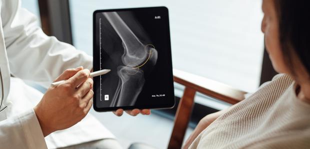
Patients on arrival will be taken care of immediately, and their condition will be diagnosed by the doctors in the orthopaedic department in conjunction with imaging services. A personalized treatment plan will then be worked out to ensure quick recovery and to regain mobility. Patients’ welfare is always a top priority.

Joint replacement surgeries too are performed here by highly qualified and experienced doctors. Patients have always been happy and satisfied with the surgical outcome.

Why Choose Us for Your Orthopaedic Care?

State-of-the-art Surgery Centre

Technology is constantly advancing and we stay up-to-date with it always. We never compromise on our equipment as we understand that our doctors’ experience and expertise is most effective and beneficial to our patients when used with the best technology and infrastructure. Our surgery centre is comparable with the best in the world and patients can rest assured that they will receive the finest care.

24×7 Comprehensive Orthopaedic Care

Day or night, patients can avail our services whenever they are in need as we are open to serve 24 hours a day, 7 days a week. At the Gleneagles Global Health City’s Orthopaedic Department, patients can always enter with the confidence that they will never be turned away.

Highest Level of Infection Control Protocols

All our staff are well-versed with the infection control protocols to be implemented and understand the need for adhering to them uncompromisingly. The fact that surgical outcomes and the patient’s welfare are dependent on the compliance to these protocols are well-entrenched in the minds of our staff and they accordingly stick to the infection control protocols with no deviation whatsoever.

Ultra-Modern Facilities

Not only are our operation theatres fitted with the best and most advanced instruments, our wards too are well-equipped and offer the patient the best comfort and care.

An Experience You Can Trust

Patients come to us apprehensive and scared, and we understand and empathize with their condition. All doubts and questions are answered with patience and we make sure the patient is well-informed about the treatment procedure that we intend to carry out.

Salient features

Complex Primary Hip and Knee Replacement Surgery

For relief from hip and knee pain due to arthritis, hip and knee replacement surgeries are suggested. Here, the hip and knee joints are replaced with implants. These surgeries are usually done for arthritic pain management in the final stages or following a fracture. We are skilled at these surgeries and patients are always happy with the mobility and the freedom they get from pain after these surgeries.

Revision Hip and Knee Replacement

While the results from primary hip and knee replacement are very good, with time, a second surgery might be needed to replace with new prostheses. This is called a revision hip/knee replacement. This procedure is more complex than a primary hip replacement and is done for the patient to regain mobility.

Oxinium (Ceramic) Total Knee Replacement

Here, oxinium (which is an alloy) is the preferred material of choice to be used as an implant in relatively young patients (50-60 Years of age), as it has increased resistance to scratching and friction is also less. With the use of an implant made of oxinium, patients get an implant that can last longer.

Complex Pelvic and Acetabular Fracture Fixation

Acute pelvic fractures are usually the result of high velocity road accidents and may be life threatening. Multi-disciplinary approach has saved many lives. Reconstruction of these fractures & acetabular fractures require special skill and training. Our doctors are experts in these.

Reverse Shoulder Replacement

For patients with massive rotator cuff injuries and those who suffer from shoulder damage due to arthritis, this surgery is suggested. As the ball and socket implants’ position is reversed, this surgery gets the name reverse shoulder replacement. We have performed this surgery successfully with satisfactory and long-lasting results.

Conditions we treat

Hip

Hip Arthritis

When patients complain of hip pain, arthritis is one of the most significant causes. With arthritis, the pain is progressive (with time the pain increases). Treatment options include physical therapy, pain killers, corticosteroid injections and surgery. The patient must refrain from high-impact activities that worsen the condition. This may become crippling if left untreated.

Osteonecrosis

Being a condition that can cause excruciating pain, it is a result of the blood supply to the femur being stopped. Cells need oxygen to survive, and in its absence they die. The hip joint, as a result, may suffer damage leading to chronic arthritis.

Hip Fractures

One of the more common hip conditions that is prevalent among the older population (above 60 years), its incidence increases further in the over 70 years age category. In older people, hip fractures can result from just a trivial fall, but the good news is if the person is otherwise healthy, with timely treatment and rehabilitation, a normal life can be restored.

Other Painful Hip Conditions

  • Bursitis: Liquid sacs called bursae are present in between tissues. Their purpose is to facilitate ease of movement and reduce friction. When this bursa gets infected, or inflamed a condition called bursitis results, which is characterized by hip pain.
  • Tendinitis: Bones and muscles are attached to each other by tendons. Due to overuse, the tendons might get sore, resulting in hip pain.
  • Muscle Strain: The hip joint is reinforced by muscles and tendons. When these are overworked, they get strained, causing hip pain.
  • Hip Labral Tear: The hip joint is cushioned by a layer of cartilage around the socket (Acetabulum). This is called the labrum. The labrum helps in keeping the thigh bone and socket together. A tear in this cartilage causes hip pain.
  • Cancer: Cancers can either originate in the hip, or they can spread to the hip from other parts. These can cause hip pain or result in fractures (Pathologic Fractures).

Knee

Knee Arthritis

The knee is the most common joint that is afflicted with arthritis. Arthritis can affect any joint in the body, causing pain and inflammation. When the knee is affected, daily activities like walking become painful. Arthritis can be controlled with proper treatment.

Fractures

In the front part of the knee, is the kneecap (patella). An injury to the kneecap in the form of an accident or a fall can cause it to crack or break. This is called a kneecap fracture and the pain from this fracture is felt immediately. Tibia and femoral fractures are major injuries and can lead to serious problems, if not attended to properly. They require immediate treatment.

Deformity Correction

Misalignment of the legs can occur due to conditions like knock knees and bow legs. In these conditions, the legs will not be straight and will look visibly deformed. The pressure borne by the knees in such cases will result in pain. Surgical correction can rectify this condition to relieve pain and give a better appearance as well.

Knee cap Pain (Patella Femoral)

Medically known as the patella femoral pain syndrome, the pain in this case can be felt anywhere around the kneecap. Being a condition common in teenaged girls, it prevents activities like running, climbing stairs, etc.,

Anterior Cruciate Ligament (ACL) Injury

The thigh bone and shin bone are connected by many ligaments. One sturdy ligament present inside the joint is called the anterior cruciate ligament. When this ligament tears (which mostly occurs during sporting activities) severe knee pain and inflammation will result. Treatment can vary as per the injury’s severity.

Other Painful Conditions and Instability

  • Meniscus Tear: The knees are cushioned by cartilages called meniscus. This cartilage can tear when a sudden force acts to twist the knee. This condition is called a meniscus tear and can be treated either conservatively or surgically depending on the injury.
  • Femoral Neuropathy: When the femoral nerve of the leg is affected by conditions like diabetes, arthritis, etc. it leads to the debilitation of the nerve. This can weaken the knees and lead to knee buckling.
  • Plica Syndrome: Plica syndrome is an inflammation of the medial plica (abnormal band of tissue in the knee) which causes knee pain and buckling.

Foot and Ankle

Fractures

Ankle fractures that are commonly encountered in young and old alike are caused by a sudden twisting movement of the foot, falls and sporting activities. Foot fractures may be caused by a fall from a height or when heavy objects are dropped on the foot. Pain while walking and inflammation are the symptoms. Treatment depends on the type and severity of fracture.

Dislocations

An ankle dislocation is usually the result of a forceful injury which causes the bones in the ankles to separate. Characterized by severe pain, visible deformation may also be observed. Diagnosed with an x-ray, the immediate line of treatment would be to restore the bones to their original position (Closed or Open reduction).

Arthritis

The feet and ankle too can be affected by arthritis. By conducting physical examinations and analysing a person’s gait, the doctor will come to a conclusion on the exact part of the foot which is affected. Proper diagnosis and treatment can cure the problem.

Deformities

The deformity may be present in the form of a flat foot (with no arch), a significantly abnormal arch, deformed toes, etc. If they do cause a problem, initially splints or orthoses with exercises help. If persistent, surgery can be performed to overcome it and restore functionality.

Other Painful Conditions

  • Bunions: Here, the big toe juts out when the tendons and ligaments do not support it properly. Being painful when the foot is fitted into narrow footwear, the condition can be overcome with orthotics and comfortable footwear. If the condition is painful, medication or surgery may be needed.
  • Achilles Tendinitis: The heel bone is connected to the calf muscles by the Achilles tendon. Overworking the tendon by running (more than what it is used to) or by performing high-impact activities injures it. Treatment involves a combination of rest, graded physiotherapy, medications and footwear modification.

Shoulder

Arthritis

Shoulder arthritis is the wear out of the cartilage inside the shoulder joint. Manifesting as shoulder pain, stiffness in the shoulder and reduced range of movement of the shoulder, it can be treated by non-invasive as well as invasive techniques. Non-invasive methods include physical therapy, icing the area, and medications.

Fracture

The scapula, the clavicle, and the humerus are the bones that make up the shoulder. Any of these can sustain a break. While they all cause shoulder pain with swelling and reduced range of shoulder movement, they also have signs unique to each of them. X-rays, CT scans, and MRIs are used to diagnose the condition. Icing the area, physiotherapy, medicines and arm slings (to immobilize the area) might help. Surgery may be needed in some type of fractures only.

Shoulder Dislocation and Instability

The shoulder joint detaches from the socket, and as a result the shoulder becomes unstable. While genetic predisposition is a cause, other causes include injury and overuse. Frequent dislocations increase instability. Patients are advised to refrain from activities that might cause dislocation along with icing and pain medications. Surgery is necessary for recurrent dislocations.

Frozen Shoulder

Medically called adhesive capsulitis, frozen shoulder is the result of the connective tissue around the shoulder thickening and reducing movement. Diabetics and people whose shoulder movement is restricted (eg. Following injury) are more prone to frozen shoulder. Physiotherapy helps along with injections to alleviate pain. In very rare cases, surgery might be needed.

Rotator Cuff Tears

The rotator cuff refers to the muscles and tendons that play a key role in connecting the upper arm with the shoulder. Symptoms of rotator cuff tear are pain at night time and pain even when the shoulder is at rest. Carrying and lifting things might also cause pain. Performing the same motion repeatedly is a common cause of rotator cuff tear. Falls, injuries, etc. are other possible causes.

Other Painful Conditions

  • Bursitis: The bursa (the sacs filled with fluid) around the shoulder that aid in movement get inflamed and cause shoulder pain.

Elbow

Fractures

The ulna is one of the bones in the forearm. The tip of the ulna (near the elbow) ends in the olecranon, which protrudes slightly and is unprotected by muscles and soft tissue. Hence, this part is susceptible to fractures and breaks/cracks relatively easily with a blow. Olecranon fracture can be treated with splint or surgery depending on the severity.

Dislocation

The radius, the ulna and the humerus form the elbow joint. When these three bones get displaced from the joint, it is called an elbow dislocation. Fall on an outstretched hand usually is the cause. Painful and visible to the naked eye, complete elbow dislocations are quite easy to diagnose. Partial dislocations will need a physical examination and diagnostic tests for confirmation.

Arthritis

Arthritis in the elbow could be osteoarthritis or rheumatoid arthritis. With osteoarthritis, reduction in the cushioning cartilage around the elbow joint causes the elbow bones to rub against each other. Injuries and overuse are the common causes.

Rheumatoid arthritis is an autoimmune condition wherein the body joints are attacked by the body’s own immune system. When both elbows are affected, rheumatoid arthritis is the usual cause.

Tennis Elbow, Golfer’s Elbow

Racquet sports like tennis, badminton, etc., which require a tight grip and swinging movement can exert a lot of pressure on the tendons on the outer part of the elbow. This condition is called a tennis elbow and it might cause pain while lifting objects, fisting the hand, raising the hand, etc. Golfer’s elbow is alike tennis elbow, except in this the tendons in the inner part of the elbow are affected. These can occur even in a person who does not play sports or games. Physical examination with imaging tests help confirm the conditions.

Other Painful Conditions

  • Cubital Tunnel Syndrome: Constriction of the ulnar nerve in the elbow can cause pain, loss of sensation and weakness in the elbow.
  • Ruptured Biceps Tendon: The biceps has tendons at both its ends with one tendon attaching to the shoulder and the other tendon attaching to the elbow. A trauma to the tendon can cause it to tear partially or completely resulting in a ruptured biceps tendon.

Hand and Wrist

Fractures

The bones of the hand and wrist may break due to road accidents, a bad fall, high-impact sports, etc. When the patient is already suffering from osteoporosis, the probability of sustaining a fracture is more. Immediate treatment is of importance as the bones might not heal properly otherwise. The symptoms include pain in the hand, swelling, and in some cases the hand is visibly deformed. A physical examination along with an x-ray is done for diagnosis. Surgery will be done under anaesthesia to realign the bones.

Arthritis

When arthritis affects the hand, performing daily activities might become difficult. Symptoms include pain which increases with hand movement. The pain is also more on waking up in the morning. As arthritis progresses, the pain also increases. X-rays, blood tests at times, are done for diagnosis. The condition’s severity as assessed on tests will determine the treatment. Treatment can be invasive or non-invasive. There are various surgical treatments too which can be done depending on individual conditions and joints involved.

Carpal Tunnel Syndrome

The median nerve runs through the arm, through the carpal tunnel in the wrist till the fingers. When this median nerve is compressed, the hands might experience a tingling sensation, or they might completely lack sensation. This is called the carpal tunnel syndrome, and it has many possible causes that range from diabetes and pregnancy to rheumatoid arthritis. What begins as a tingling sensation may aggravate to pain and the inability to hold objects properly. Early treatment is important for recovery. Treatment options include immobilizing the hand, performing stretching exercises, pain medication, and surgery as the last resort.

Trigger Finger

When bending the finger, the finger gets locked in bent position and straightens with an audible snap accompanied by pain. This is called trigger finger. Overuse of the fingers and hand, performing the same movement repeatedly and inflammation of the tendons can result in trigger finger. Pain on getting up in the mornings, pain while gripping objects and straightening fingers are the other symptoms. There are no specific tools to diagnose this condition; a physical examination is what will help the doctor in diagnosis. Rest, immobilization, physiotherapy, and pain medications will help treat the condition. Surgery when done for unresponsive individuals gives dramatic relief.

Other Painful Conditions

  • Cubital Tunnel Syndrome: When the ulnar nerve is pinched, cubital tunnel syndrome might result. When the elbow is subjected to a lot of weight, this condition manifests. Surgical and non-surgical treatment options are available.

Musculoskeletal Tumors

Benign Tumours of the Bone

Just like in other parts of the body, tumours can form in the bones and muscles too. These tumours can be benign or malignant.

Benign tumours occur in any age. The tumour may be felt as a lump and may also be accompanied by pain. Sometimes, its presence is discovered accidentally while taking an x-ray for a fracture. The fracture may have been a result of the tumour’s growth. It is best to surgically remove the tumour.

Bone Metastasis

In bone metastasis, cancer from other parts of the body spreads to the bones. The cancer does not originate in the bone. When the cancer affects the bones, it can form small holes in the bone. As a result, the bone gets weakened. This is called osteolysis, and the possibility of fracture increases when bones are thus affected. Radiotherapy, chemotherapy, pain management and surgery are the treatment options.

Others

Pelvic and Acetabular Fractures

Usually sustained in an accident or because of a fall from height, pelvic and acetabular fractures require prompt treatment. For senior citizens though, even a relatively insignificant fall can result in this fracture if their bones are already weak. The complicated anatomy of the pelvis makes this a challenging condition to treat. The pelvis holds up the upper body, and the acetabulum refers to the part of the pelvis that connects to the femur. These fractures usually are accompanied by injury to the surrounding tissues and nerves. The surgeon will decide on a surgical or non-surgical option depending on individual conditions.

Bone Infection (Osteomyelitis)

When the bone is infected, either as a result of infection spreading from far away tissue or by infection originating in the bone itself, the condition is called osteomyelitis. In the latter case, a wound or open injury might expose the bone to germs and result in the condition. Fever with pain and inflammation at the site of infection are the usual symptoms. Smokers and diabetics are at an increased risk of contracting this condition, and hence they should take care to avoid cuts in feet, which may lead to osteomyelitis.

Geriatric Orthopaedics – Fragility Fracture

When a fracture is sustained by a fall from standing height or less, this is usually because the bones are already weak. This kind of fracture is called a fragility fracture and is common among older citizens. With a fragility fracture, surgical intervention is recommended so that the patient can regain mobility as quickly as possible. Quick recovery is all the more important as in some cases immobility can lead to various complications. Post-surgery, methods to prevent future fractures need to be implemented.

Periarticular Fractures

Fractures that occur in or in close proximity to a joint are called periarticular fractures. Extreme pain and inability to move are characteristic of these fractures. Treatment is complicated because of the anatomical nature of these fractures. Though they can occur anywhere in the body, parts of the leg are more common sites for these fractures. Since they are close to the joints, accurate treatment is necessary.

Periprosthetic Fractures

In a periprosthetic fracture, the bone breaks or cracks near the implant that has been placed already. These types of fractures are becoming increasingly common these days. They might be caused by weak bones, bone infection, and pressure on the bone. Women are more at risk than men.

Post-Surgical Problem Fractures (Malunion and Non-union)

In a malunion, the bone heals after a fracture, but its positioning is not right. As a result, the limb may seem twisted/ bent or reduced in length. When pain persists after fracture treatment, malunion is a possible cause. Surgical intervention may be required to correct it.

Non-union refers to the fractured part’s inability to mend. Even for months after the fracture, healing does not take place. For a fracture to heal well, it needs to be held in proper position with adequate blood supply. When blood supply is not enough, non-union can result.

Orthobiologics

Orthobiologics are substances that are found in the human body that promote healing. So, surgeons make use of these to enhance the healing process. Bone grafts, stem cells, platelet rich plasma, etc. are all examples of orthobiologics.

Orthopaedic Procedures

Joint Replacement

  1. Primary Total Hip Replacement

  2. Here, the parts of the hip that are damaged are replaced by artificial implants. Usually, total hip replacement is suggested when arthritis has completely damaged the hip joints. When the patient is unable to lead a normal life because of hip pain, this procedure will aid in restoring near normalcy. The arthritis could be due to various causes.

    • Osteoarthritis: Here, the cartilage inside the joints is damaged. Hence there is more friction and consequently more wear and tear.
    • Rheumatoid Arthritis: This is an autoimmune condition in which the body’s immune system attacks its tissues.

    Other than arthritis, osteonecrosis might also lead to a need for total hip replacement. Preparing for surgery involves giving a detailed medical history to your doctor, performing various blood tests and getting an x-ray. The surgical procedure is performed under general or spinal anaesthesia. Post-surgery, you might have to stay at the hospital 3 to 4 days. Smoking should be avoided as it inhibits healing. Physiotherapy will be started at the hospital, and this has to continue even after the patient goes home. Physiotherapy is an important part of the recovery process as it aids in strengthening the area. In 6-12 weeks, most of the daily activities can be resumed. However, high-impact sports should be avoided. Your doctor will guide you on the exercises that can be carried out without stressing the joint.

  3. Revision Total Hip Replacement

  4. A revision hip replacement might be needed by patients who have undergone total hip replacement in the past. The reason could be wear and tear of the prostheses, after which it becomes painful. Another reason could be an infection which damages the areas surrounding the hip joint. Both these conditions will cause a lot of hip pain and will inhibit normal functioning and movement. To restore mobility and improve quality of life, revision hip replacement is done.

    Surgical preparation involves blood tests, x-ray, bone scans, etc. The doctor will need a detailed list of the medications the patient is taking; especially if the patient is on aspirin or blood thinners, the doctor has to be kept informed. During surgery, the old prostheses in the hip are replaced with new ones. Sometimes, if the hip is infected, more than one revision surgery will be required. In the first, the prosthesis will be removed. After the infection has been treated and healed, a second surgery will be performed to place a new prosthesis. 5 -7 days of hospitalization will be required. Recovery involves regular physiotherapy. Recovery is longer than after primary hip replacement and may take 6 weeks to 3 months.

  5. Primary Total Knee Replacement

  6. When the knee is damaged due to arthritic changes and even simple daily activities cause pain, a total knee replacement is suggested to relieve pain and perform normal activities once again. Pain while walking, climbing stairs, sitting down and getting up, etc. might all be successfully relieved with this procedure. The surgeon will perform a few tests to make sure the knee replacement will benefit the patient. These include tests to check the knee’s range of movement and strength. The choice of prostheses is varied, and the surgeon will choose one after taking into consideration the patient’s age, the activity the knee will be subjected to, knee structure, etc.

    The surgery will take around 1 1⁄2 – 2 hours and will be performed under general or spinal anaesthesia. Hospitalization of around 4-5 days will be required. For the post-surgery pain, medications will be given. On the same day following surgery, physiotherapy will commence. The patient will also be apprised on how to take care of the surgical site and what diet to consume. If high fever sets in or if the patient experiences rigor or inflammation in the knee area, the doctor should be consulted immediately. Recovery will take around 3-6 weeks. Activities that do not stress the knees can be performed. High-impact sports are best avoided.

  7. Revision Total Knee Replacement

  8. Total knee replacements are very successful surgeries. But like everything else, the prosthesis used in total knee replacement can wear out. After that, it will need to be replaced. Factors like body weight and high-impact activities will also have an effect on the life of the prosthesis. Thus, a prosthesis that was once firmly fixed might move, leading to knee pain. Also, minute particles may get eroded from the plastic spacers used in the previous surgery, and these particles will face action from the body’s immune system. During the process, the bones too might get attacked by the immune system leading to a condition called osteolysis. This again will require revision knee replacement. Infection is a possibility in any surgery. An infection may set in immediately or even many years after the primary total knee replacement. The patient will experience pain, rigidity and inflamma-tion in the knee. This is another condition which will make revision surgery a necessity.

    A revision surgery is more complex than the initial primary total knee replacement. Talk to the doctor about the possible complications of surgery. A number of tests will be conducted to determine the feasibility of surgery. Done under spinal anaesthesia, the procedure will take 2-3 hours. The surgeon will remove the old implant and replace it with a new one. Bone grafting might be done as well. After surgery, hospitalization of 5 days will be needed. After discharge, the patient will need help at home too for the ini-tial few days.

  9. Partial Knee Replacement

  10. When arthritis has damaged only a part of the knee, surgery can be done to replace the damaged part alone. This is called partial knee replacement. Done under either spinal anaesthesia, a prosthesis replaces the damaged part only. Recovery is relatively quicker. The range of motion in the knee after surgery is also more. Complete recovery will take around 2-3 weeks

  11. Total Shoulder Replacement

  12. Arthritis may cause severe shoulder pain in some patients. Movement of shoulder without experiencing pain may not be possible, thus debilitating the patient. To get pain relief, total shoulder replacement is performed. Another reason to consider total shoulder replacement is a bad fracture.

    In total shoulder replacement, shoulder joints are replaced with prostheses. Before surgery, diagnostic tests like x-ray or MRI will be done. Blood tests too will be conducted. The surgery may be performed under general anaesthesia, with the patient in a seated position. The joints affected by arthritis will be replaced with prosthesis. After surgery, the arm movement will be restricted with a sling. Gradual movements will be started. Recovery will take around 6- 8 weeks. Lifting heavy objects should be avoided.

  13. Reverse Shoulder Replacement

  14. The shoulder joint is indeed one of the more complex joints in the human body. The upper arm bone and the shoulder blade meet to form the ball and socket joint of the shoulder. In a reverse shoulder replacement, as the name implies, the position of the ball and socket are interchanged. To elaborate, a metal ball takes the place of the socket and a plastic socket replaces the ball. This makes the joint more stable, and there is no need for the surrounding tendons to provide stability. Thus, in cases where the tendons are also damaged, reverse shoulder replacement is a good option. Patients who suffer from arthritis along with a torn rotator cuff are ideal candidates.

    The surgical procedure will take around 3 hours. The patient will have to stay in the hospital for 4-5 days. This surgery is not recommended for patients who smoke as it will inhibit recovery. It is also not recommended for patients with systemic infection and those suffering from severe osteoporosis.

  15. Total Elbow Replacement

  16. The elbow joint is a hinge joint where three bones meet. The bones are the humerus, the radius, and the ulna. There is a cartilage present at the point where the bones meet each other to aid in smooth movement. There is also a synovial membrane which secretes a fluid to help in friction-free movement. The elbow joint is stabilised by the surrounding muscles and ligaments. When the elbow is damaged and the pain prevents normal activities, total elbow replacement is done wherein the elbow joints are replaced with prosthesis.

    The reasons for elbow damage could be many: rheumatoid arthritis, osteoarthritis, a bad road traffic accident, etc. Depending on the extent of damage, total or partial elbow replacement may be required. The surgery will be performed under general anaesthesia and will last around 3 hours. Before surgery, a number of investigations will be done and detailed medical history will be recorded. Any medications the patient is on should be communicated to the doctor. After surgery, hospitalization of 2-4 days will be needed, during which antibiotics will be given to prevent possible infection. Physiotherapy will also be taught. Total elbow replacement will help the patient resume normal activities. However, activities that involve a lot of pressure on the elbows are best avoided.

Joint Preservation

  1. Core Decompression of the Hip and Stem Cell Therapy

  2. For young patients with early stages of osteonecrosis, core decompression is the preferred treatment. The procedure involves drilling being done into the dead bone tissue. Drilling has many benefits: it reduces swelling, allows better flow of blood, and inhibits further damage to the bones. The success of the procedure is dependent on how much of the bone has already suffered damage, which part of the bone has been damaged, the amount of weight, the stress the joint is subjected to, etc. An MRI done ahead of the surgery will tell the surgeon exactly which bone tissue needs removal. This removal will stimulate the production of healthy bone tissue.

    The bone marrow that is present inside the bones contains stem cells. These stem cells can change into bone cells when they are injected into the body. This stem cell therapy is used along with core decompression for a better surgical outcome.

  3. High Tibial Osteotomy

  4. A high tibial osteotomy is typically recommended for younger patients suffering from arthritis of knee, to prevent the need for a total knee replacement. When only a part of the knee has suffered damage and not the entire knee, this procedure would be a good choice. During the procedure, a wedge is made in the upper part of the tibia. This helps to remove the weight from the damaged tissue and shift it to healthier tissue. This procedure serves to delay the need for a total knee replacement by at least 10 years.

    An x-ray will be done before the surgery to ensure that osteotomy is the right choice and to determine the plan for the surgery. Performed under spinal anaesthesia, the surgery will last for around 2 hours. Hospitalization of two to three days may be required. Walking aids should be used after surgery so that the knee is protected from stress. Physiotherapy after surgery is a must and should be performed diligently for best results.

  5. Osteochondral Autograft Transfer System (OATS)

  6. The knee joints in our body are covered by cartilage. The purpose of the cartilage is to ensure that the joint movement is smooth and free from friction. Sometimes this cartilage can suffer injury because of high-impact activity. The patient will experience pain, inflammation, and inability to move the joint freely. To address this, healthy cartilage from the part of the knee that doesn’t bear much weight replaces the damaged cartilage. Performed as an arthroscopic surgery, it is minimally invasive. The surgeon removes the damaged cartilage and replaces it with the new one.

    Before surgery, smoking should be avoided and the surgeon kept informed about all the drugs the patient is on. Talk to the surgeon about your doubts and get them clarified. The surgery will be done under spinal anaesthesia. The rehabilitation after surgery is very important and the surgeon and his team will guide the patient through it.

  7. Bone Marrow Aspirate Concentrate (BMAC)

  8. BMAC injections are helpful in providing pain relief from osteoarthritis. The injections contain stem cells that are harvested from the patient’s own bone marrow. When injected into the part that has been affected by osteoarthritis, the patient gets sufficient pain relief.

    The procedure takes around an hour to an hour and a half and is performed under spinal anaesthesia. The stem cells are first harvested from the patient’s pelvis. It is processed and then injected back into the damaged area that is then covered in bandages. There may be some pain after the procedure. The doctor will guide the patient on managing it. It is to be noted that some painkillers are not allowed after this procedure. Pain relief after the procedure will take around 6 weeks. The pain relief will last for quite some time, and there usually is no need to repeat the procedure at short intervals. BMAC is not recommended for patients suffering from cancer, tuberculosis, radiotherapy, etc. The patient should communicate any condition they may be suffering from to the doctor so that the doctor can take an informed decision about giving the injection.

Arthroscopy

Knee

  1. Anterior Cruciate Ligament Reconstruction

  2. The anterior cruciate ligament is an important ligament in the knee that acts as a bridge between the bone of the thigh and the shin. The ligament traverses the knee in the middle and stabilizes it. When this ligament tears (which happens mostly during high-impact contact sports), the person will feel severe pain and buckling of the knee. In such cases, ACL reconstruction can be performed to replace the ligament with a tendon. The tendon is usually taken from patients same or from opposite leg.

    Before surgery, physical therapy will commence with the goal of reducing inflammation. This is done to improve the outcome of surgery as a stiff, rigid knee may not benefit fully from the surgery. The procedure will be performed under spinal anaesthesia through very small openings or slits. After the procedure, the patient will have to use crutches with the knee being secured in a brace for protection. The doctor will help in pain management after surgery. Detailed post-operative care instructions will be given, which will have to be followed. Physiotherapy too will start. Recovery will take a few weeks.

  3. Multi-ligamentous Injury Knee Reconstruction

  4. In a multi-ligament knee injury, two or more of the knee ligaments suffer damage. Research has shown that treatment with surgery gives better outcome than any other non-invasive method. However, the surgery is a complicated one and needs detailed physical examination and imaging tests like x-ray and MRI. Sometimes, more than one surgery may be necessary. The surgery done arthroscopically is least invasive. The exact procedure will differ from patient to patient – for some patients, only one ligament may need repair, while for another all three might need reconstruction. Typically, grafts from the patient or a donor will be used for reconstruction.

    After surgery, it is important to slowly mobilize the knee while keeping the reconstructed ligaments intact. Mobilizing the knee early will give the knee a better range of movement. Crutches and knee braces can be used. Depending on the patient’s progress, the doctor will take a call on when to walk without crutches. Knee strengthening and other physiotherapy exercises should be carried out for the next 4-6 months after surgery. The doctor will schedule reviews at regular intervals to monitor the patient’s progress.

  5. Meniscectomy

  6. The knee contains two menisci – one on the exterior edge of the knee and the other towards the interior. These menisci perform many vital functions:

    • They help in smooth, friction-free movement of the knee.
    • They help in distributing the body weight over the knee joint.
    • They absorb shocks and sudden jerks.
    • They provide stability to the knee joint.

    Damage to the menisci may lead to the need for either a total or partial meniscectomy. Menisci injuries are quite common and minor tears will not need surgery. But if the menisci do not mend with conservative treatments or if the knee starts to lock, surgery is recommended. When a surgery is decided on, it is best to start knee-strengthening exercises before the surgery to improve surgical outcome. The surgery itself, done arthroscopically, will involve only three small cuts to repair the menisci. During surgery, the damaged parts of the menisci are removed. Patients are usually discharged on the same day. They can walk but will have to use crutches to prevent over-exerting the knee. Mild pain and inflammation in the knee are expected after surgery and are nothing to be concerned about. Pain medication will be given to manage it. It is important to do strengthening exercises at home after discharge.

  7. Meniscal Repair

  8. Meniscal repair is another surgery, also done arthroscopically, to repair torn menisci in the knee. Usually sustained during some kind of sporting activity, meniscal tears are accompanied by a popping sound. Walking after the tear might be possible but painful. Meniscal tears may also be caused by wear and tear, but this kind of tear usually subsides on its own without surgical intervention. Other symptoms that indicate meniscus tear are inflammation, limited movement of knee and a feeling of instability in the knee. Diagnosis involves the McMurray test and imaging tests to confirm diagnosis. If non-invasive treatments are ineffective, then surgical meniscal repair is performed.

    During surgery, the torn menisci is sutured and then held in place. Physiotherapy will commence after surgery and complete recovery will take 4-6 weeks.

  9. Root Repair

  10. In a meniscus root tear, the part of the meniscus that attaches to the bone i.e. the root of the meniscus is damaged. The pain from a meniscus root tear is usually chronic; the tear itself can limit knee movement, make the knee unstable and increase the risk of osteoarthritis. The root tear can be of two types: traumatic and degenerative. A traumatic tear usually affects sportspeople and hence is more common in younger adults. A degenerative root tear is caused as a result of wear and tear; older adults are more prone to this. When the doctor is of the opinion that non-surgical treatments will not yield desired results, surgery is done arthroscopically.

    During surgery, the roots of the meniscus are repaired and connected to the bone once again. Recovery after surgery will take around 4-6 weeks. During the recovery period, the patient will be advised to use crutches to minimize the load on the knees; physiotherapy will also commence.

  11. Cyst Excision

  12. A cyst in the knee, called a popliteal cyst or a Baker’s cyst, may develop in people suffering from arthritis. Usually found at the back of the knee, the cyst is in simple terms a sac of fluid. Surgery can be done to remove the cyst, but it is important to understand that the cyst may recur unless the underlying cause is treated.

    The knee joint is covered in cartilage. The synovial membrane, which is the lining of the joint, produces a fluid to keep the joints moving smoothly. When an excess of this fluid is produced, the extra is contained in a sac at the back of the knee in the form of a cyst. This cyst can be felt when the back of the knee is touched. Other than arthritis, a knee injury may also be a cause of the cyst.

    Treatment can be surgical or conservative. Only if the cyst is very painful, surgery is recommended. During surgery, the cyst is removed and any damage caused by the cyst to the lining of the joint is rectified. Treating the cause of the cyst is important to prevent its recurrence.

  13. Cartilage Defect Treatment -OATS, BMAC

  14. As the name suggests, a cartilage defect is a part of the cartilage that has been injured. The cause may be accidents, osteonecrosis or other knee conditions. Cartilage defects usually occur along with damage to knee ligaments like an ACL tear. The surgeon will always try to treat the defect non-invasively with physiotherapy, medication and injections. Only if this is not successful will other invasive procedures be considered. With a cartilage defect, the patient may experience pain and also a possibility of arthritic changes. Diagnosis is done with an MRI.

    • Treatment with OATS – Osteochondral autograft transfer is preferred for younger adults especially, if the bone is also damaged. Here, healthy cartilage replaces the defective cartilage.
    • Treatment with bone marrow aspirate concentrate (BMAC) – This procedure is carried out when the affected area is large. Stem cells from the bone marrow are harvested and injected back into the patient.

Shoulder

  1. Bankart’s Repair

  2. Like in all other joints, there is cartilage surrounding the shoulder socket as well. This cartilage, called the glenoid labrum, can suffer damage due to sporting injuries or other shoulder conditions. This tear to the cartilage is called Bankart’s tear and is quite a common condition. Shoulder pain is the symptom, and if left untreated, shoulder dislocation may become more frequent. Treatment starts with conservative methods; if the desired results are not obtained, the doctor will suggest the Bankart’s repair, which is done arthroscopically.

    The surgery involves small incisions. During surgery, the cartilage and any torn ligaments are reconnected to the bone. The recovery duration after surgery will vary from patient to patient – it can be anywhere between 3-6 months. Physical therapy plays a very important role in strengthening the shoulder and restoring range of movement.

  3. Latarjet Procedure

  4. The shoulder has three bones which all join to create a ball and socket joint. The ball sits in the socket of the shoulder and is held in that position by various ligaments and tendons. When these ligaments and tendons are not able to keep the ball in the socket, shoulder dislocation takes place. Left untreated, repeated dislocations could damage the bone of the shoulder blade. A shoulder dislocation could manifest as severe shoulder pain, swelling, and inability to move the shoulder normally. The Latarjet procedure is recommended for such patients having recurrent dislocations, especially in those with a bony defect. Earlier the surgery was done as an open surgery, but now it can be done through arthroscopic means.

    During the procedure, the coracoid from the shoulder blade replaces the damaged bone and thus prevents further dislocations from happening. The success rate of the procedure is good, and recovery will take around 3-6 months. The shoulder will be immobilized with the help of a sling during recovery. Physical therapy is important, and complete recovery can be achieved in around 6 months.

  5. Rotator Cuff Repair

  6. The rotator cuff in the shoulder is made of muscles and tendons. These muscles and tendons secure the ball of the shoulder in its socket. Thus, it is responsible for us being able to perform a number of functions like raising our hands up and being able to do activities like swimming, basketball, etc. Simply put, the ability to rotate our shoulder is attributed to the rotator cuff. A rotator cuff tear may happen due to overuse of the shoulder. Other causes include road traffic accidents, lifting big objects and a fall with your body weight being taken up by the arm.

    To treat rotator cuff repair, earlier an open surgery had to be performed. But now, it is possible to do the procedure arthroscopically. The procedure differs depending on the kind of tear and is decided on by the surgeon. As in any procedure, rehabilitation plays an important role in recovery. Physiotherapy is started with mild exercises and will progress to exercises involving more movement. Recovery takes around 6 months. The results after surgery are encouraging with patients reporting pain-free shoulders with increased stability.

  7. Neglected Rotator Cuff Tear Repair

  8. A rotator cuff tear will not heal without treatment. Supply of blood is necessary for healing, and in its absence, the rotator cuff tear will worsen. The initial line of treatment is always conservative – in the form of physiotherapy. There are no medicines available to heal a torn rotator cuff; medicines are given only to alleviate pain. In physiotherapy, other muscles and tendons are strengthened to make up for the function of the torn rotator cuff. Unfortunately, there is always a chance of the rotator tear getting inflamed again.

    When timely treatment is not given, the tear can get bigger, and the shoulder joint can become extremely unstable. This condition is called cuff tear arthropathy. Sometimes a condition called pseudo-paralysis may manifest where the range of shoulder movement is badly affected. Both are serious conditions that need aggressive treatment. For cuff tear arthropathy in senior citizens, a reverse shoulder replacement is recommended. If the patient is younger, then a muscle transfer (taking a muscle from the shoulder to do the function of the rotator cuff) or a superior reconstruction (introducing a graft from the thigh to aid in shoulder stability) can be done.

  9. Subacromial Decompression

  10. When the roof of the shoulder presses down on the rotator cuff, it results in a condition called shoulder impingement that is very painful. In most cases, it is a bone spur that presses the rotator cuff. This condition only aggravates with time. It starts as mild shoulder pain; the pain may be more when the hand is raised. Slowly the pain increases and lifting the hand overhead may not be possible at all. As with any treatment, surgery is the last option and is considered only when physiotherapy, medications, and corticosteroid injections do not give the desired results.

    The surgical procedure is called subacromial decompression or acromioplasty. The surgery is done arthroscopically through small cuts. It involves removing the tissue and, in some cases, removing the bursa too if it is inflamed. The entire procedure is performed under general anaesthesia. Recovery typically happens within a month with the shoulder being immobilized in a sling. Once the sling is removed, physiotherapy will commence and complete recovery will take a year.

Complex Trauma

  1. Pelvic – Acetabular Trauma

  2. Pelvic-acetabular fractures are often very complicated and are usually caused by a road traffic accident. To treat these fractures, sometimes more than one surgery might be necessary. In patients suffering from osteoporosis, even a fall might cause this fracture.

    Many bones make up the pelvis which, other than supporting the upper body, also protects the intestines and bladder. When one of the pelvic bones suffers a break, it is called a pelvic fracture. The joining point of the pelvis with the femur is called the acetabulum, which is a ball and socket joint. When the fracture is in the socket, it is called an acetabular fracture. An acetabular fracture is difficult to treat because of its position close to major organs and important blood vessels.

    During surgery, the bones are positioned properly and then held by screws and plates, etc. Pain and complications after surgery will be treated by the surgeon and his team. The patient will be encouraged to not remain immobile as immobility may lead to complications. If many nerves are damaged, recovery may take from 6 months to more than a year.

  3. Intraarticular & Comminuted Fractures

  4. When a fracture occurs across a joint, it is called an intraarticular fracture. Such fractures are very complex as they involve bones inside the joints unlike fractures involving only bones that are relatively simpler to treat. The condition could be made more complex by bone fragments, which will slow down healing. Mostly caused by road traffic accidents, these fractures usually affect the wrist, the elbow, the shoulder, the hip, the knee and the ankle. The patient will experience immediate pain, inflammation and inability to move the affected part. Treatment starts with immobilizing the injured area. The surgeon will decide on open surgery or an arthroscopic procedure. During surgery, the bones will be repositioned and held in place by screws, wires, etc. The doctor will guide the patient on post-operative care and rehabilitation.

    A comminuted fracture is one in which the bone has been broken at three or more places. It is further classified as open and closed. In an open comminuted fracture, the skin is cut, while in a closed comminuted fracture there is no visible injury to the skin. Sustained as a result of an accident or bad fall, diagnosis involves x-ray, CT, MRI, etc. Surgery involves repositioning the bones and holding them in place with rods, plates and screws, etc. Following surgery, immobilization with casts and pain management with medications will begin. In some cases, traction might also be needed. For an open fracture, the doctor will also take care to clean the wound to prevent infection.

  5. Osteotomies and Realignment Surgeries for Malunion Fractures

  6. A malunion happens when a fracture heals but with bones positioned improperly. The symptoms of a malunion are pain, rigidity of the part, limited movement, and in some cases the bone looks visibly deformed. It is necessary to treat a malunion as a failure to do so may cause arthritis, instability, and ultimately loss in functionality. Diagnosed with imaging tests, a malunion can be corrected through surgery. A procedure called osteotomy is done to realign the bone. During surgery, depending on individual conditions, the surgeon may shorten the bone and reposition it or may use a graft to fix the malunion. Surgery will be followed by physiotherapy to increase range of movement of affected part. This surgery can restore functionality of affected part and also prevent deterioration in bone health.

  7. Bone Grafting & Revision Surgeries for Fracture Non-Unions

  8. In normal circumstances, after surgery, the bones will heal with time and new tissue will form and reconnect. In some cases, however, even when a surgical procedure is carried out successfully, the fracture might not mend/join. This condition is called a non-union. The reasons for non-union vary from diabetes and hypothyroidism to lack of healthy food and old age. Another possible cause is the lack of blood supply needed for the fracture to heal.

    The doctor may suggest a bone graft to treat a non-union. The graft could be taken from another part of the patient’s body or harvested from another individual or a synthetic graft. When the graft is introduced, it launches new bone cells and chemicals needed for healing to take place. In addition to a bone graft, metal plates and screws will be used to hold the bones in place.

Foot and Ankle Surgery
  1. Ankle Arthrodesis

  2. Ankle arthrodesis is a surgery that involves fusing the bones of the ankle to overcome the effects of arthritis. The ankle has two joints:

    • The tibiotalar joint that is the meeting of the shin bone with the foot bone.
    • The subtalar joint formed when two of the tarsal bones in the foot meet.

    Arthritis can lead to pain and swelling in the aforementioned joints. Ankle arthrodesis done to treat this can be performed either as an open surgery or arthroscopically. During surgery, the joints will be pressed together and then secured in that position using metal screws, nails, etc. Sometimes, a bone graft may be used to hasten healing. Post-surgery pain is expected. It will be managed with medication; keeping the foot raised will also help. The foot will be immobilized in a brace to prevent any movement. Complete recovery will take 3-6 months.

  3. Lisfranc Fracture

  4. A Lisfranc fracture sustained as a result of contact sports, fall, or road traffic accidents, involves the bones in the mid-section of the foot. The Lisfranc ligament is found in between two bones (medial cuneiform and second metatarsal) of the foot. These two bones meet to form the Lisfranc joint. Any of the aforementioned injuries can damage the bones and injure the ligament. Pain felt in the middle of the foot, inflammation and inability to exert weight on the foot are the symptoms of this condition. Diagnosed with a physical examination and imaging tests, your doctor might suggest surgery to correct it.

    During surgery, the bones will be realigned and held in place with metal plates, screws, wires etc. Any ligament injury will also be attended to. Alternatively, the surgeon might also perform a fusion surgery. Here, the bones are fused together. Both procedures will need foot immobilization for a few weeks.

Paediatric orthopaedics

  1. Club Foot

  2. Club foot is a congenital condition in which the baby’s foot will be turned towards the inside and will also possibly face downwards. It mostly affects both the feet, but the new-born will be otherwise healthy. In club foot, the tendons of the foot will be shorter than usual. For severe club foot, surgery is done to correct it, and doctors recommend early surgery so that the baby’s walk is not affected. Parents however need not feel concerned that the baby will find club foot painful. Though it looks painful, club foot does not hurt the baby.

    Club foot can be treated with casts and braces. A small surgery to the Achilles tendon might also be carried out. Wearing the brace properly according to the doctor’s instructions plays a very important role in recovery.

    Alternatively, for severe club feet, surgery might be done to stretch the tendons and ligaments to position the foot properly. Here again, the use of casts and braces will be needed for best results.

  3. Perthes Disease

  4. Perthes disease is a rare condition that affects certain children. Here, the blood supply to the top of the thigh bone is impaired. When the bone does not get enough blood, the bone cells die (necrosis).

    The thigh bone, or the femur, ends in a ball that fits into the socket of the pelvis. As a result of the reduction in blood supply to the bone, the top of the thigh bone (the ball) may weaken and ultimately collapse. The exact cause for this condition is unknown, though there is a probability that the condition might be genetic. Children in the age group of 4-10 years are the ones usually affected.

    When the child is affected with Perthes disease, there will be a noticeable change in the way he/she walks and runs. Hip pain that increases with activity and a limp may be the other symptoms. Diagnosed with a physical examination (in which the doctor will check the child’s ability to move the legs) and an x-ray, the doctor may suggest treatment options depending on the individual child’s condition.

    Non-surgical options include not stressing the legs too much with sporting activities, physiotherapy and using casts.

    Surgery is recommended if the damage to the top of the thigh bone is bad. An osteotomy is usually performed wherein the bones are cut and realigned. They are made to fit well within the socket and held in position with metals screws, plates, etc. Post-operative care involves the use of crutches, casts, and rehabilitation exercises.

  5. Slipped Capital Femoral Epiphysis

  6. This is a condition that can manifest in adolescents. In adults, the thigh bone ends in a ball that fits into the socket of the acetabulum, thus forming the ball and socket joint. In children who are still growing, the head of the femur is called as the epiphysis. This epiphysis and the rest of the thigh bone are connected by a growth plate. In a slipped capital femoral epiphysis, this interconnecting growth plate is injured and the Epiphysis is displaced. Reasons for this may vary from obesity and hypothyroidism to endocrine problems. Boys are more susceptible to the condition compared to girls. Leg pain, reduced range of movement in the leg, change in walking style, etc. are the possible symptoms. It is usually diagnosed with a physical examination and an x-ray; in some cases an MRI may be required.

    The condition is treated with surgery. A metal screw is used to join the femur head to the femoral bone. If the bones have been badly damaged, the surgeon may have to reposition the bones. After surgery, the use of crutches will be necessary to protect the leg till it heals.

  7. Cubitus Varus/ Genu Valgus Deformities

  8. Cubitus Varus Deformity

    Cubitus varus, also known as gunstock deformity, is a condition in which when the arm is extended; the palm is facing front, and the arm is turned towards the body. Fracture malunions may result in this condition. Very rarely, it is present as a congenital defect. Sometimes, infections, tumours and bone cell death might also cause this condition. If the deformity does not cause any trouble, doctors may just decide to observe the condition. If the ulnar nerve is compressed, then surgery might be needed. The surgeon will perform an osteotomy to realign the elbow and treat the condition.

    Genu Valgum Deformity

    A genu valgum deformity is the opposite of a varus deformity i.e., the limb is turned away from the body at the knee. Knee pain may be a result of this deformity, and the uneven pressure distribution might result in arthritic changes in the knee. It can be corrected with surgery during which the bones are repositioned.

Others

  1. Limb Reconstruction Surgeries for Tumour Conditions

  2. Tumours can be malignant or benign. They can either originate in the bone itself, or they may spread to the bone from the other parts of the body. Diagnosing a tumour will involve blood tests, x-ray, and possibly a biopsy. When the tumour is found to be benign, unless it is causing a problem, the doctor may decide to just observe it. If it is found to be pressing on a nerve or blood vessel, then surgery to remove it may be suggested.

    For malignancy, surgery is suggested either before or after chemotherapy. The exact procedure will differ from patient to patient depending on their condition. Earlier, if the tumour had spread badly, then amputation was being done. Now with the development in medical technology, it is possible to carry out extensive reconstruction procedures to save the limb.

  3. Minimally Invasive Plate Osteosynthesis (MIPO)

  4. Due to road traffic accidents, fractures might happen at three or more places in the bones (comminuted fracture). Treating this (while being minimally invasive) is done with a procedure called minimally invasive plate osteosynthesis. In a comminuted fracture, along with the breakage in bone, bone fragments may also be present. This makes treatment all the more difficult. To avoid the disadvantages that are present in more conventional treatment methods, MIPO is done to reduce the possibility of infection as well as malunion and non-union. The other advantages of MIPO include:

    • Reduction in the time taken for surgery
    • Loss of blood is minimized
    • Soft tissue harm is kept to a minimum
    • Reduction in swelling
    • Lesser recovery time
  5. Debridement, Sequestrectomy, & Antibiotic Instillation

  6. Debridement or sequestrectomy is done to accelerate healing in tissues in osteomyelitis. When a wound does not get better, removing the dead cells might start the healing process. Debridement reduces the possibility of infection and promotes growth of new, healthy cells. Debridement is done under anaesthesia, wherein the surgeon removes the dead tissue and (if need arises) puts a graft in place. When the procedure is done in conjunction with antibiotic instillation, the results are much better than when the procedure is done alone.

Best Ortho Doctors in Chennai

Gleneagles Global Health City, Chennai provides you with one of the best Orthopaedic doctors and Orthopaedic surgeons in the country. Our Orthopaedic consultants are widely renowned in their respective fields and come with a wide range of experience and expertise. Specialists in various branches of Orthopaedics, our doctors deliver comprehensive treatment and care. Visit us at Gleneagles Global Health City, for consulting with the best team of Orthopaedic doctors and Orthopaedic surgeons in Chennai.

Best Orthopaedic Surgeon Chennai DrKesavanRajagopalanAmruthur
Dr Kesavan A R

M.B.B.S, M.S

Senior Consultant – Department of Orthopedics & Joint Replacement

Dr Sivakumar A P
Dr A P Siva Kumar

M.B.B.S, M.S (Ortho)

Consultant – Department of Orthopaedics & Joint Replacement

Dr Shiva Reddy
Dr Shiva Reddy

M.B.B.S, D.N.B

Consultant, Department of Orthopedics and Joint Replacement

Related Blogs & Videos

 
Ganglion Cyst
Everything you need to know about ganglion cyst

Know More

Tennis Elbow
Everything you need to know about tennis elbow

Know More

Frozen Shoulder Min
Frozen shoulder Treatment in Chennai

Know More

Related Blogs & Videos

 
We are with you in your journey to better health

A consultation with our panel of doctors, specialists and surgeons will help you determine what kind of services you may need to help diagnose and treat your condition. If you or someone in your family or friend’s circle are facing any health issues, please get in touch with us, we are here for you.