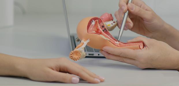
Urology – Overview

What is Urology?

Urology is the branch of surgery which deals with the Male Urinary and Reproductive tract and Female Urinary tract. The Urinary tract consists of the Kidneys, ureters, Urinary Bladder, Urethra. The Male Reproductive organ consists of the Penis, Testes, Vas deferens, Prostate and Seminal vesicles. Kidney stones, prostate enlargement, cancers of the urinary tract, male sexual dysfunction, stress incontinence in women, urinary tract infections are some of the common problems dealt by an urologist. Kidney transplantation is another area where the urologist plays a key role. Diseases of the adrenal gland also come under the purview of the urologist.

Department Overview

The department of Urology at the Gleneagles Global Health City (GGHC) has some of the best urologists in the country. The department specialises in comprehensive urological treatment of a variety of illnesses affecting the urinary tract. The department consists of highly skilled urologists committed to the best quality patient care. They are supported by equally efficient technical and nursing staff. The department specialises in the treatment of the entire urinary tract, including the kidneys, adrenal glands, bladder, urethra, prostate, seminal vesicles and penis. Our doctors are equipped to deal with patients in a professional and sensitive manner, ensuring patient comfort and speedy recovery.

Sub-Speciality Services

Renal Transplantation

The kidneys perform the vital function of filtering the blood and removing wastes and toxins from the system. If both the kidneys stop working, the subsequent build up of wastes and toxins in the body can cause other complications. Renal or kidney transplantation is usually only done in patients who are suffering from end stage kidney disease who are dependent on dialysis for their excretory function. During the procedure the kidney which is retrieved either from a living donor or a deceased (brain dead) donor is implanted into the patient’s abdomen. The patient’s diseased kidneys are not usually removed.

The kidney for the transplantation may be donated by a deceased donor or a living donor, such as a family member. The donor kidney is carefully chosen based on the patient’s blood type and human leukocyte antigen type (HLA). This helps to minimise the chances of the body rejecting the donor organ. Rejection occurs when the body’s immune system identifies the donor kidney as a foreign body and starts to produce an immune response to attack it. The patient is prescribed immune suppressant medications after the surgery to lower the risk of organ rejection.

Uro-Oncology

Uro-oncology is one of the areas or sub-specialities in urology concerned with the diagnosis and treatment of cancers of the urinary tract. Uro-oncology also deals with cancers of the male reproductive organs. Kidney, bladder and prostate cancer are the most common urologic cancers. Our uro-oncologists treat malignant growths in the urinary tract with a variety of tools at our disposal including surgical intervention, radiation therapy and chemotherapy. Surgical intervention may be laparoscopic or open. The Uro-oncologist with the collaboration of the medical oncologist tailors the treatment according to the individual patient to deliver the highest standard of care.

Endo-Urology

Endo-urology is a sub-specialty within the field of urology which uses minimally invasive surgical techniques used to treat diseases of the urinary tract. It is commonly used in the diagnosis and treatment of stone diseases of the urinary tract. During the procedure, a highly sophisticated endoscope is passed into the patient’s natural urinary passage to access the stone. The stones are then fragmented using a laser. Some of the common endo-urological procedures performed are URS (uretero-renoscopy), RIRS (Retrograde Intra-Renal Surgery). Large kidney stones can be removed by a procedure called as PCNL (Per-Cutaneous Nephro Lithotomy) wherein the kidney stones are accessed by a small keyhole incision through the back. Endo-urology is a real boon to the patients as it eliminates the need for a large surgical incision and allows the patient to recover faster.

Reconstructive Urology

Reconstructive urology is another area in urology which aims to restore normal anatomy and function to the genitourinary tract. This includes bladder reconstruction, urethral reconstruction for strictures, pelvic floor reconstruction. Traumatic injuries to the genitourinary tract or congenital defects may also require reconstructive surgery. Reconstructive surgeries are unique because they require a high degree of customisation to match the patient’ needs.

Laparoscopic procedures for cancers of the urinary tract

Traditional open procedures to remove tumours of the urinary tract require large incisions and lengthy hospital stay. With advances in medical science and the evolution of laparoscopic techniques, it is possible to treat these diseases through keyhole incisions. During a laparoscopic procedure, the surgeon inserts a laparoscope (a small tube with a camera) and specialised instruments through small incisions in the abdomen. The camera provides enhanced visualisation of the tumour, which enables the surgeon to remove the tumour without much blood loss. Post operative pain and scar are less in laparoscopic procedures compared to open procedures. Even large tumours can be removed safely using laparoscopic techniques.

Female Urology

Female urology is a sub-specialty of urology that deals with treatment of urological conditions affecting women. A female urologist deals with problems like female urinary incontinence, pelvic organ prolapse, over-active bladder, under-active bladder, interstitial cystitis etc. Most of these procedures are done by minimally invasive techniques.

Paediatric Urology

Paediatric Urology is another sub-specialty focussing on diseases of the genito-urinary system of children. It is a highly specialised field. Some of the problems dealt in this sub-speciality are Pelvi-ureteric junction obstruction (PUJO), Hypospadias, kidney stones in children, phimosis, neurogenic bladder, undescended testis, wilms tumour, bladder exstrophy etc.

Male infertility and Erectile Dysfunction (Andrology)

Intracytoplasmic sperm injection (ICSI) is an In-vitro fertilisation (IVF) procedure in which a single sperm is injected into an ovum. This procedure is most effective for couples with severe male infertility. Typically, men with severe infertility do not have many sperms in their ejaculate, so sperm retrieval is done directly from the epididymis or the testes. Depending on the cause of the male infertility, different methods may be chosen to extract the sperm like testicular sperm aspiration (TESA), percutaneous epididymal sperm aspiration (PESA), testicular sperm extraction (TESE) or micro-epididymal sperm aspiration (MESA).

Erectile Dysfunction is another common problem dealt by an urologist. The patient is subjected to a comprehensive evaluation with the help of blood tests including hormonal assays and imaging studies like penile Doppler. Penile prosthesis is typically recommended for severe cases of erectile dysfunction where all other options have failed.

Neuro-Urology

Neurological conditions affecting bladder can cause problems like difficulty in voiding (under-active bladder) or leakage of urine (overactive bladder). Patients suffering from spinal cord injuries, Parkinsons disease, stroke, Alzheimer’s disease, diabetes will have variable degrees of neurogenic bladder. Complete evaluation including urodynamic study is performed and appropriate treatment instituted.

Why Choose Us?

Patient Experience

At GGHC, Patient Experience is our highest metric for quality. We know how important it is for the patient to be comfortable while they heal, and we do our best to ensure it. The doctors and nurses monitor the patients continuously and update their treatment protocols as required. Our ultimate aim is always to ensure the patient has a good quality of life after treatment. From admission to discharge and post-treatment follow-ups, our doctors take great pains to educate the patient and make sure they are comfortable.

best urology treatment center india

Latest Technologies

GGHC is known for world-class healthcare with a compassionate touch. We embrace technology and believe in investing in the best infrastructure to help us give our patients the best chances of a full recovery. The hospital has streamlined procedures and diagnostics to minimise overlaps and unnecessary waste of time. Our highly advanced diagnostic and treatment facilities are among the best in India.

best urology treatment center india

Providing Quality Care

The doctors in various departments work together and provide high quality inter-disciplinary care to all our patients. We pride ourselves at being able to treat complex diseases of the urinary tract and associated organs. Our doctors are assisted by an efficient nursing and technical staff, to ensure the patients are monitored closely and cared for.

best urology treatment center india

Procedures

Cystectomy

A cystectomy is a surgical procedure in which a portion of the bladder is removed. Cystectomies may be performed to treat recurrent invasive or non-invasive bladder cancer. Cystectomies may also be performed to treat advanced colon, prostate or endometrial cancer, interstitial cysts or congenital abnormalities. Radical cystectomies in men involve removing the seminal vesicles and prostate in addition to the bladder. In women, radical cystectomies result in the removal of the bladder, the uterus, the ovaries and a part of the vagina. Cystectomies can be performed via an open procedure or using minimally invasive techniques.

The cystectomy procedure also deals with reconstruction of an alternate channel to divert and store urine in, after the bladder is removed. This can be done in a number of ways. The surgeon may harvest a small piece of the small intestine to make an ileal conduit. Neobladder reconstruction using a larger piece of the small intestine is also an option.

Pyeloplasty

Pyeloplasty is a surgical reconstruction technique for the ureter. It also helps drain and decompress the kidneys. The procedure is used to relieve an obstruction in the ureteropelvic junction. Pyeloplasty procedures can be performed laparoscopically or as an open procedure in teenagers and adults. In infants and small children, an open pyeloplasty is considered safer. The pyeloplasty procedure has the highest success rate in treating kidney and ureter obstructions. There is evidence of a good long-term prognosis for patients who have undergone a pyeloplasty.

During a pyeloplasty, the obstructed segment of the ureter is removed. The remaining ureter is then attached to the renal pelvis (on the kidney). A stent may also be placed to drain urine from the kidney and relieve pressure. Risks associated with the procedure include scarring, herniation, infection and blood clots.

Uroflowmetry

Uroflowmetry is a diagnostic screening procedure to calculate the patient’s urine output over time. The test is non-invasive and used to assess bladder and urinary sphincter function. During the uroflowmetry test, the patient is asked to urinate into a funnel until their bladder is completely empty. The funnel is connected to a measurement apparatus that measures urine flow rate with respect to time and converts the data into graphs. During normal urine output, the initial stream starts slowly, speeding up until the bladder is nearly empty. Then, the last part of the bladder empties slowly. The shape of the flow rate graph will vary if there is an obstruction, and the degree of variation will give the doctor information about the size and location of the obstruction in the urinary tract. Decreased flow rate could also be an indication of urinary incontinence caused by weak bladder muscles. The doctor may require a series of uroflowmetry tests over the course of multiple days. Uroflowmetry tests are used in conjunction with imaging tests to diagnose urological diseases like bladder cancer, urinary obstructions and enlarged prostate gland.

Urodynamic study

Urodynamic tests are used to measure urological nerve and muscle function, bladder pressure and urine flow rates. The tests essentially study how well the bladder, urethra and sphincters operate while holding and releasing urine. If the patient suffers from frequent painful urination, incontinence or problems with emptying the bladder completely, the doctor may recommend urodynamic tests to figure out the underlying problem. Types of urodynamic tests include cystometry, electromyography, uroflowmetry, pressure flow studies and postvoid residual measurements. A cystometric test measures bladder pressure via a catheter inserted up the urethra. Electromyography uses sensors to measure electrical activity of the bladder muscles and sphincters. Postvoid residual studies measure the urine that remains in the bladder after urination with an ultrasound.

Renal Stents

Renal artery stenting is a procedure used to unblock an obstructed renal artery, the main blood vessel that carries blood to the kidney. Renal arteries can get blocked due to atherosclerosis or fibrous artery disease. Blockage of the renal artery reduces blood flow and oxygen supply to the kidneys, resulting in deteriorated functioning. When the kidneys stop filtering the patient’s blood, it can lead to dangerous levels of toxins and wastes building up in the blood, poisoning the body and increasing renal pressure. A renal stent is inserted into the blocked artery to restore blood supply to the kidney.

Renal stents are inserted during a surgical procedure known as an angioplasty. This is a minimally invasive procedure, in which a balloon catheter is inserted into the renal obstruction through a small incision in the skin. The surgeon guides the tip of the balloon catheter to the precise site of the obstruction under X-ray fluoroscopy guidance. The surgeon will inject contrast dye to make the arteries easier to visualise, before inflating the balloon. The stent, made of a fine mesh tube, is inserted into the narrowed artery. It is left in place to maintain a path for blood flow. The patient will require periodic follow-up appointments to check if the artery is narrowing again.

Trans-Urethral Resection of the Prostate (TURP)

The prostate gland, found only in men, is located close to the bladder. Inflammation of the prostate gland or benign prostate hyperplasia can press on the bladder and the urethra causing discomfort and difficulty while urinating. Trans-urethral resection of the prostate is a minimally invasive surgical procedure to remove the enlarged prostate through the penis. The surgeon accesses the enlarged prostate by inserting an instrument through the urethra. The instrument is called a resectoscope and is a long tube with a camera on the end, valves that control irrigation fluid and a wire loop that can be used to make cuts and cauterize. The surgeon uses the wire loop to resect pieces of tissue blocking the urethra and flushes it out through the bladder with the help of the irrigation fluid. After the trans-urethral resection, a catheter is left in place for a few days as blood and residual tissue continues to be flushed out through the urine.

Prostate Biopsy

A prostate biopsy is a procedure to retrieve tissue samples from a suspicious mass in the prostate. The sample is tested further in the laboratory to identify the nature and cause of the abnormal mass. During the procedure, the urologist inserts a needle into the prostate gland to collect a tissue sample. The procedure is quick and only lasts about 10-15 minutes. The tissue sample is examined under a microscope to identify cell mutations in the mass to identify a probable cause and a course of treatment. A doctor may recommend a prostate biopsy if the patient has lumps during a rectal exam or elevated prostate-specific antigen (PSA). Prostate biopsies are associated with risks such as rectal bleeding, blood in the semen or the urine and in rare cases, infection.

Flexible Cystoscopy

A flexible cystoscopy is a procedure used to examine the inner lining of the ureters and the bladder. The flexible cystoscope, a hollow tube with a camera on the end, is slowly inserted into the urethra and advanced upwards into the bladder. The cystoscopy may be performed in an examination room, using a local anaesthetic jelly to numb the area. It may also be performed as an outpatient procedure under local anaesthesia or a surgical procedure under general anaesthesia. The attending physician will decide on the type of procedure depending on the condition of the patient. Flexible cystoscopy procedures could be used to diagnose, monitor or treat conditions affecting the bladder. The doctor may recommend a flexible cystoscopy to patients who present with blood in the urine, incontinence, painful urination or chronic urinary tract infections.

Intravesical Chemotherapy for bladder cancer

Intravesical chemotherapy is a technique in which chemotherapy is delivered directly to the bladder. Intravesical chemotherapy is effective in treating non-invasive tumours that do not spread beyond the bladder. Normally, chemotherapy is injected into the blood and allowed to work. This affects other parts of the body as well. In intravesical chemotherapy, the effects of the chemotherapy target the inner lining of the bladder, minimising side-effects in other parts of the body. Intravesical chemotherapy is often recommended after trans-urethral resection of bladder tumours, to kill any residual cancer cells. Experts typically recommend that the chemotherapy be administered within 6 to 24 hours of the tumour resection procedure, but the surgeon in charge of the case may decide the time based on the condition of the patient. The main side effects of intravesical chemotherapy for bladder cancers are blood in the urine and a residual burning sensation in the bladder.

Double J stent removal

A double J stent is a hollow plastic wire inserted in the ureter connecting the kidney to the bladder. The stent ensures a flow of urine from the kidney to the bladder without obstructions. An obstruction could cause renal hypertension (high pressure in the kidney), acute discomfort or pain during urination. The double J stent needs to be removed periodically due to the material of which it is made – flexible plastic. The stent needs to be removed within the specified time or else it could lead to complications in which the stent disintegrates and begins to pass through the patient’s urinary tract in pieces, potentially blocking the ureters and leading to infections. The double J stent is removed in a cystoscopy procedure. The cystoscope is inserted into the urethra, while the area is numbed with an anaesthetic jelly. Once the stent is visualised via the camera on the cystoscope, it is removed using endoscopic forceps. The entire procedure usually takes about 30 minutes. Mild burning sensations in the urethra and some blood in the urine are common after a cystoscopy procedure.

Penile Prosthesis implantation

A penile prosthesis is a device implanted in the penis to treat erectile dysfunction. The penile prosthesis is typically recommended only after other treatment options for erectile dysfunction have failed. The implant devices are usually malleable or inflatable. The inflatable implant is more advanced and generally preferred by most patients. The penile prosthesis is implanted in a surgical procedure. The recovery time for the surgery is usually around 6 weeks but varies according to the patient’s general health, the cause of the dysfunction or defect, the type of implant and reconstruction techniques involved.

Kidney preserving procedures

Kidney tumours discovered at an early stage have a good chance of curative treatment without removing the entire kidney. Partial kidney resection is recommended for small, non-aggressive tumours. This can be done through a partial nephrectomy. This is a laparoscopic procedure in which the tumour and surrounding margins are resected and removed laparoscopically. This allows preservation of the normal anatomy and function of most of the organ.

Diseases and Treatments

Anterior Prolapse

Anterior Prolapse Overview

Anterior prolapse (known as anterior vaginal prolapse in women) or prolapsed bladder occurs when the bladder descends from its position and pushes on the vaginal walls or the pelvic floor muscles. This happens when the pelvic floor becomes weak or if sudden pressure is put on them, commonly during childbirth or heavy lifting. Mild to moderate cases of anterior prolapse can be treated non-surgically. In more severe cases, surgical intervention may be required to repair the prolapse.

Signs and symptoms of Anterior Prolapse

Mild cases of anterior prolapse may not present with any symptoms at all. If symptoms occur, they may include pressure in the pelvis or vagina, especially while coughing or lifting heavy objects and strain while urinating. Patients may have a hard time maintaining a steady stream of urine or struggle with urinary incontinence. Symptoms may appear to go away when you lie down and become more pronounced while standing for long periods of time. The patient may be susceptible to frequent urinary tract infections.

Causes of Anterior Prolapse

Anterior prolapse occurs when the bladder sinks due to weakness or trauma to the pelvic floor muscles. The bladder sinks and causes a bulge in the vaginal wall. The stress to the pelvic floor could be caused by childbirth, heavy lifting, repeatedly strained bowel movements, violent coughing or being overweight.

Risk factors of Anterior Prolapse

Women who are overweight, pregnant women who have undergone multiple deliveries and post-menopausal women are at a higher risk of developing anterior vaginal prolapse. Having your uterus removed in a hysterectomy procedure could also weaken the pelvic floor muscles and increase your risk of an anterior prolapse, but that is not always the case.

Complications of Anterior Prolapse

An anterior vaginal prolapse can lead to urinary incontinence and significant pain during sexual intercourse.

Diagnosis of Anterior Prolapse

An anterior vaginal prolapse is diagnosed on the basis of a patient’s medical history and a physical examination. The doctor may also require the patient to undergo a urodynamics test to measure the bladder’s ability to hold and release urine and a cystourethroscopy to examine the urethra and bladder for blockages.

Treatment of Anterior Prolapse

Anterior prolapse may not require surgical treatment unless it is symptomatic. Mild cases can be treated with pelvic floor muscle exercises or a pessary. A pessary is a plastic or rubber ring inserted into the vagina to support the prolapse and relieve pain. A moderate to severe anterior prolapse will require reconstructive surgery to move the bladder into position. Post-surgical recovery usually takes four to six weeks.

Prevention of Anterior Prolapse

People at a high risk of developing anterior prolapse should refrain from heavy physical work and maintain a healthy body-mass index.

Bladder Cancer

Overview of Bladder Cancer

Bladder cancer is a common type of cancer that originates in the cells of the bladder. The cancer usually begins in the urothelial cells which comprise the inner lining of the bladder. Most bladder cancers are discovered and diagnosed at an early stage. At this stage, the disease is easy to treat. However, bladder cancer can recur, so even patients who have been rid of the cancer need to have periodic follow-ups.

Signs and symptoms of Bladder Cancer

The symptoms of bladder cancer include blood in the urine, frequent urination, painful urination and back pain. In some cases, the blood in the urine may be microscopic and only detectable in urine tests.

Causes of Bladder Cancer

Bladder cancer is caused by abnormal cells in the bladder that begin to multiply at an alarming rate. This is usually the result of a cell mutation. The abnormal cells multiply and form a tumour that can grow and spread. Bladder cancer can metastasise, i.e., it can spread to other parts of the body. Bladder cancer is classified into various types based on the type of tissue cells in which the cancer originates – Urothelial carcinoma (inner bladder lining), Squamous cell carcinoma and adenocarcinoma.

Risk factors of Bladder Cancer

Family history of bladder cancer, a smoking habit and previous cancer treatments can increase a patient’s risk of developing bladder cancer. Repeated bladder infections can also increase a patient’s risk of bladder cancer. Men of an advanced age appear to be the most high-risk demographic. Exposure to certain heavy industrial chemicals like arsenic and lead can also increase a person’s risk of developing bladder cancer.

Complications of Bladder Cancer

Advanced bladder cancer can be fatal.

Diagnosis of Bladder Cancer

Tests and procedures used to diagnose bladder cancer include a cystoscopy to visualise the tumour endoscopically, a biopsy of the tumour, urine cytology and a series of imaging tests including a CT scan and X-ray scans.

Treatment of Bladder Cancer

The treatment for bladder cancer is customised according to the patient and the stage the cancer has progressed to. The treatment program could include a combination of surgery to remove the tumour, chemotherapy, radiation therapy and immunotherapy.

Prevention of Bladder Cancer

There is no 100% effective method to prevent bladder cancer. However, minimising risk factors can help reduce the chances of getting bladder cancer.

Bladder Stones

Overview of Bladder Stones

Bladder stones are hard lumps of mineral build-up that occur in the bladder. Bladder stones may occur when minerals in concentrated urine crystallise in the bladder into lumps. This can be caused due to chronic dehydration resulting in a high concentration of minerals in the urine and failure to empty the bladder completely. Bladder stones are most common in men above the age of 50.

Signs and symptoms of Bladder Stones

Small bladder stones may not cause symptoms. Common symptoms associated with bladder stones are abdominal pain, blood in the urine, burning sensation, difficulty during urination and cloudy urine.

Causes of Bladder Stones

Bladder stones are formed when the bladder does not empty fully. A number of conditions could prevent complete emptying of the bladder including a swollen prostate, nerve damage to the bladder or the urinary sphincter, bladder diverticula and anterior vaginal prolapse. A diet high in fats, salts and sugar can also accelerate the formation of bladder stones.

Risk factors of Bladder Stones

Men over the age of 50 are the demographic most vulnerable to bladder stones.

Complications of Bladder Stones

Bladder stones that do not pass can cause urinary tract blockages that lead to infections.

Diagnosis of Bladder Stones

To diagnose the patient, the doctor will begin with a physical exam and a personal and family medical history. The doctor will also require a panel of urine tests, a cystoscopy (an endoscopic exam to look for blockages in the urinary tract) and imaging tests including a CT scan, an ultrasound and an X-ray scan.

Treatment of Bladder Stones

Small bladder stones can pass through on their own. The patient may be advised to drink a lot of water to help the stone pass. If they do not pass on their own, the doctor may prescribe medication to break up the stones and ease their passage. Larger bladder stones may require surgical intervention. The surgeon may choose to perform an open extraction procedure or use endoscopic lasers to break up the stones and flush them out.

Prevention of Bladder Stones

To prevent bladder stones, drink plenty of water and focus on emptying the entire bladder during every urination.

Urinary tract infection (UTI)

Overview of Urinary tract infection (UTI)

Cystitis is a medical condition characterised by an inflammation of the bladder. In most cases, cystitis is caused by a bacterial urinary tract infection. Cystitis could also occur when the body’s naturally occurring bacterial ecosystem becomes imbalanced. Cystitis may be triggered by certain medications or personal hygiene products. Long-term use of a catheter for urination could also cause cystitis. The treatment for cystitis usually depends on the underlying cause. Cystitis can occur in anyone but is most common in women.

Signs and symptoms of Urinary tract infection (UTI)

Cystitis can cause many uncomfortable symptoms including a frequent urge to urinate, burning sensation during urination, blood in the urine, cloudy or smelly urine, pelvic discomfort and low-grade fever. If the patient experiences extreme symptoms such as nausea, vomiting, fever, chills and back pain, they should be rushed to the hospital immediately.

Causes of Urinary tract infection (UTI)

Cystitis is usually caused by a UTI, which is a bacterial infection. However, it could also be caused by certain medications including chemotherapy, radiation therapy, long-term use of urinary catheters and irritating personal hygiene products.

Risk factors of Urinary tract infection (UTI)

Women are at a higher risk of developing cystitis than men. Pregnant women, women who use diaphragms for birth control and post-menopausal women are at the highest risk of cystitis.

Complications of Urinary tract infection (UTI)

Cystitis rarely causes serious complications when treated. However, chronic untreated cystitis can cause kidney infections and haematuria (blood in the urine).

Diagnosis of Urinary tract infection (UTI)

The diagnosis of cystitis begins with a physical exam and a patient medical history. The doctor will also require a urine analysis, a urine culture, a cystoscopy to visualise the bladder and a set of imaging tests including CT scans, ultrasound scans and MRI scans.

Treatment of Urinary tract infection (UTI)

The treatment of cystitis depends on the exact cause. Bacterial cystitis is treated with antibiotics, fluids and anti-bacterial vaginal creams. For mild cases of cystitis, the patient can ease their symptoms at home with fluids, heating pads and over-the-counter painkillers. Cystitis triggered by certain products can be reversed by avoiding those products.

Prevention of Urinary tract infection (UTI)

Taking plenty of water, emptying the bladder completely while urinating, wiping from front to back after bowel movements and avoiding deodorant sprays in the genital area are also recommended preventative measures.

Hematuria

Overview of Haematuria

Haematuria refers to the presence of blood in the urine. Visible blood in the urine is called gross haematuria. Blood in the urine that is only visible under microscopic testing is known as microscopic haematuria. It is critical to identify the cause of the bleeding. The course of treatment is based entirely on the cause.

Signs and symptoms of Haematuria

Gross haematuria causes red coloured urine. This is due to the presence of red blood cells in the urine. The bleeding is usually not painful unless the patient is passing blood clots. Certain foods such as beetroot, rhubarb and berries can also turn the urine pinkish, but this usually stops within a few days.

Causes of Haematuria

Haematuria could be caused by urinary tract infections, kidney infections, enlarged prostate, sickle cell anaemia or internal bleeding due to kidney trauma. Haematuria could also be caused by advanced kidney, bladder or prostate cancer.

Risk factors of Haematuria

Anyone can develop haematuria but it is most common in men above the age of 50, who have enlarged prostate glands. A recent kidney or bladder infection, a family history of haematuria and certain medications like aspirin and penicillin can increase your risk of haematuria.

Complications of Haematuria

Haematuria could be an indication of cancer. Untreated haematuria could lead to kidney failure.

Diagnosis of Haematuria

Haematuria is diagnosed on the basis of a physical exam, a patient’s medical history, detailed urine analysis and imaging tests including a CT scan and an MRI scan. The doctor may also require a cystoscopy, wherein an endoscopic camera is inserted into the urethra and bladder to examine for causes of the haematuria. Sometimes the cause of the haematuria remains undiagnosed.

Treatment of Haematuria

The treatment for haematuria depends on the cause. Haematuria caused by urinary tract infections may be treated with antibiotics. The doctor will prescribe the appropriate medication in case an enlarged prostate is causing the haematuria. Kidney or bladder stones may be treated with shock therapy. If it is due to cancer, appropriate treatment will be recommended. The patient will have to follow up with the doctor to ensure that presence of blood in the urine is eliminated after the course of treatment.

Prevention of Haematuria

To prevent haematuria, the patient can take measures to prevent the underlying causes such as kidney or bladder stones, urinary tract infections and enlarged prostate gland.

Overactive Bladder

Overview of Overactive Bladder

Overactive bladder, also called OAB, is a bladder condition in which the patient may feel a sudden, frequent urge to urinate uncontrollably. The patient may have a disturbed sleep because of this. The condition can have a debilitating effect on the patient’s social and professional life. Luckily, OAB can be treated.

Signs and symptoms

A patient with OAB may feel a sudden, uncontrollable urge to urinate very frequently. They may also experience incontinence during these episodes. Typically, this disrupts the patients sleep and ability to lead a normal life.

Causes of Overactive Bladder

OAB is caused by an involuntary muscle contraction of the pelvic floor muscles and other muscles that operate during urination. Several conditions can contribute to OAB including diabetes, neurological disorders like stroke, urinary tract infections, menopause and bladder abnormalities including bladder stones.

Risk factors of Overactive Bladder

Patients with enlarged prostate glands and diabetes are at a higher risk for OAB. Patients with Alzheimer’s disease and other neurological diseases are also at a higher risk for OAB.

Complications of Overactive Bladder

OAB can cause long-term mental health problems like anxiety and depression, due to the social trauma of the condition. The patient may also suffer from sleep-deprivation and issues with sexuality.

Diagnosis of Overactive Bladder

The diagnosis for OAB begins with a patient medical history, an account of their symptoms and a pelvic or rectal exam. The doctor will also require a panel of urine tests and neurological tests.

Treatment of Overactive Bladder

The treatment for OAB is usually a combination of approaches. The doctor may recommend a range of behavioural therapies including Kegel exercises, biofeedback therapy, intermittent catheterization and scheduled toilet breaks. The doctor may also recommend medications, bladder injections or nerve stimulants in combination with the behavioural therapy. If these treatment methods are ineffective, the doctor may recommend surgical intervention.

Prevention of Overactive Bladder

Healthy lifestyle choice can help decrease your risk of OAB – Maintain a healthy weight and get regular exercise. Drink a lot of water. Limit alcohol, smoking and caffeine intake. Practicing Kegel exercises to tighten the pelvic floor muscles can also help.

Urinary Incontinence

Overview of Urinary Incontinence

Urinary incontinence is the involuntary leakage of urine from the bladder. This could be caused by loss of control or weakness in the urinary sphincter. Urinary incontinence is a condition that affects many people. It affects more women than men. Urinary incontinence could be caused by many different factors. Urinary incontinence can have an adverse effect on the patient’s lifestyle. If you have urinary incontinence, please do not hesitate to seek medical help.

Signs and symptoms of Urinary Incontinence

Many people experience an occasional, involuntary release of urine. However, when patients experience incontinence regularly, it becomes a problem that needs to be treated. The patient may experience incontinence during moments of stress or when sneezing or coughing. Some patients experience a constant trickle of urine due to a bladder that does not empty completely.

Causes of Urinary Incontinence

Urinary incontinence has a long list of possible causes. Urinary incontinence could be caused by pregnancy, vaginal childbirth, menopause, enlarged prostate, prostate cancer, any urinary tract obstruction, multiple sclerosis, Parkinson’s disease, stroke, brain tumour or a spinal injury.

Risk factors of Urinary Incontinence

Being overweight, smoking and having neurological disorders can increase your risk of urinary incontinence. Women are more likely to develop urinary incontinence than men.

Complications of Urinary Incontinence

Chronic urinary incontinence can cause urinary tract infections, skin rashes and affect the patient’s mental health.

Diagnosis of Urinary Incontinence

The doctor begins diagnosing the patient with a medical history and a physical exam (pelvic and rectal). The doctor will require a full urinalysis to assess the possible causes. The patient will also have to maintain a bladder diary and have a post-void urine measurement study.

Treatment of Urinary Incontinence

There are a range of treatment options available for urinary incontinence. The doctor usually starts with less-invasive methods such as behavioural therapy and medication. This may include bladder training, Kegel exercises, double-voiding and fluid management. If these are ineffective, the doctor may suggest a urethral insert or a pessary to provide relief. The doctor may recommend surgery in case none of these methods work for the patient.

Prevention of Urinary Incontinence

Urinary incontinence is not entirely preventable. However, quitting smoking, having a healthy diet, limiting alcohol and caffeine can help reduce the risk. Practicing Kegel exercises to keep the pelvic floor muscles strong is also recommended.

Prostate Cancer

Overview of Prostate Cancer

Prostate cancer is an uncontrolled growth and division of mutated cells in the prostate gland. The prostate gland is a walnut-shaped gland that produces seminal fluid, an important component of the male ejaculate. Prostate cancer is very common and tends to grow slowly. Prostate cancer usually does not metastasise to other organs. However, there are aggressive variants of prostate cancer too.

Signs and symptoms of Prostate Cancer

Prostate cancer may not cause any symptoms in the early stages. As the cancer progresses to a more advanced stage, the patient may experience decreased force in the stream of urine, difficulty in urinating and blood in the urine and semen. The patient may also experience bone pain, have erectile dysfunction and unexplained weight loss.

Causes of Prostate Cancer

Prostate cancer, like most other cancers, is caused by a genetic mutation which causes uncontrolled cell division. The exact cause or origin of the gene mutation that causes prostate cancer is unknown though.

Risk factors of Prostate Cancer

Men above the age of 50 are at the highest risk of prostate cancer. Family history and obesity could also be strong risk factors for prostate cancer.

Complications of Prostate Cancer

Prostate cancer can, in some cases, metastasise to other organs. Prostate cancer and its treatment could result in urinary incontinence or erectile dysfunction.

Diagnosis of Prostate Cancer

The requirement for regular screening tests for prostate cancer in healthy men with no symptoms is controversial, but it is important for men with a family history of the disease. Prostate cancer screening includes a Prostate-specific antigen test and a digital rectal exam. If the screening tests detect a prostate abnormality, the doctor may require an ultra-sound scan, and MRI scan and a prostate biopsy to confirm the diagnosis of prostate cancer. The aggressiveness of the tumour is then gauged using the Gleeson score and genomic testing.

Treatment of Prostate Cancer

The treatment for prostate cancer depends on the stage at which it is discovered and its aggressiveness. Early-stage, slow-growing tumours may not require immediate treatment. The doctor may elect to actively survey the tumour, keeping a periodic check on its growth and metastasis. Surgery to remove the prostate is an effective option for advanced prostate cancer. This can be done laparoscopically or through an open procedure.

Prevention of Prostate Cancer

Prostate cancer cannot be prevented, but the risk can be reduced by certain healthy lifestyle habits. This includes having a healthy diet, exercising regularly, maintaining a healthy weight and having regular prostate exams. If you have a family history of prostate cancer, consider getting routine screening tests for prostate cancer.

Our Doctors

Dr Muruganandham K
Dr Muruganandham K

MBBS, MS, DNB, MCh, FMAS

HOD & Senior Consultant – Department of Urology

Dr Karthik V C
Dr Karthik V C

M.B.B.S, M.S, M.Ch

Consultant

Related Blogs & Videos

 
We are with you in your journey to better health

A consultation with our panel of doctors, specialists and surgeons will help you determine what kind of services you may need to help diagnose and treat your condition. If you or someone in your family or friend’s circle are facing any health issues, please get in touch with us, we are here for you.